
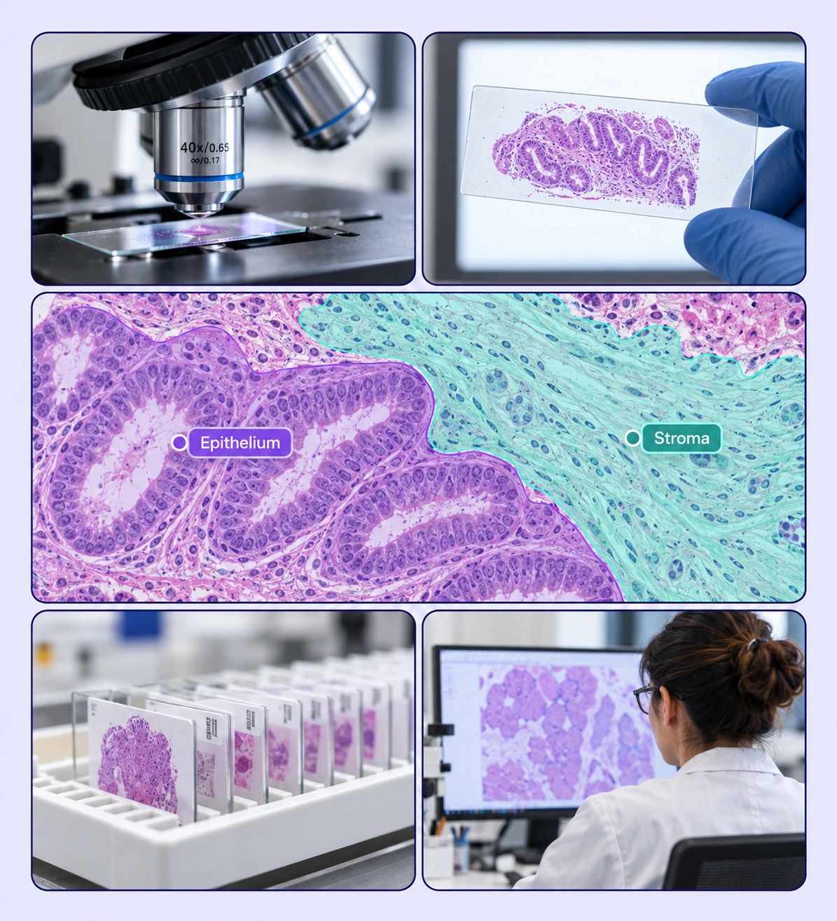
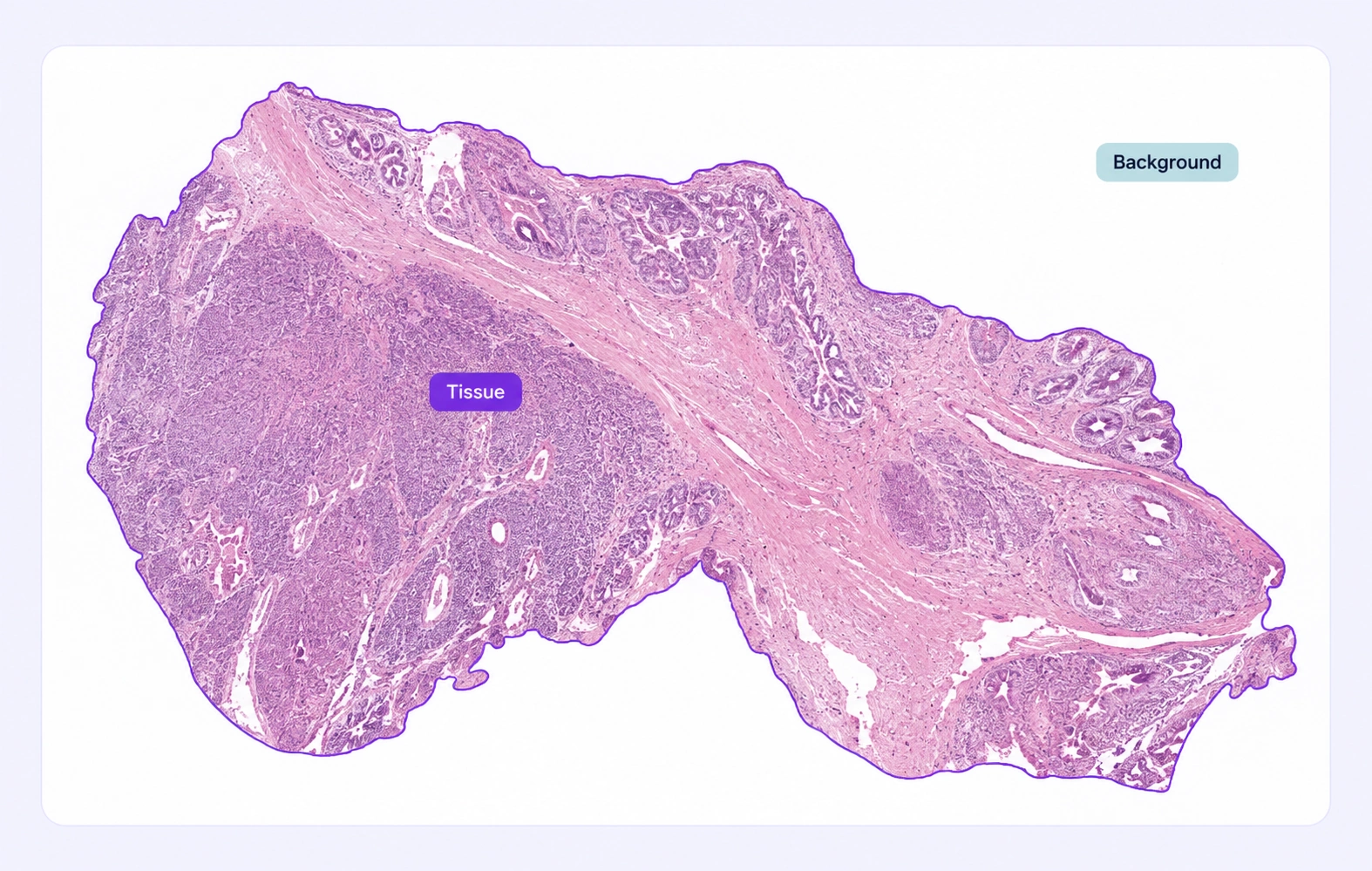
Segment tissue regions with model-assisted labels and expert review workflows.
Automate curation, management, and versioning of tissue datasets.
Reusable ontologies and governed quality control reduce labeling and review rework.
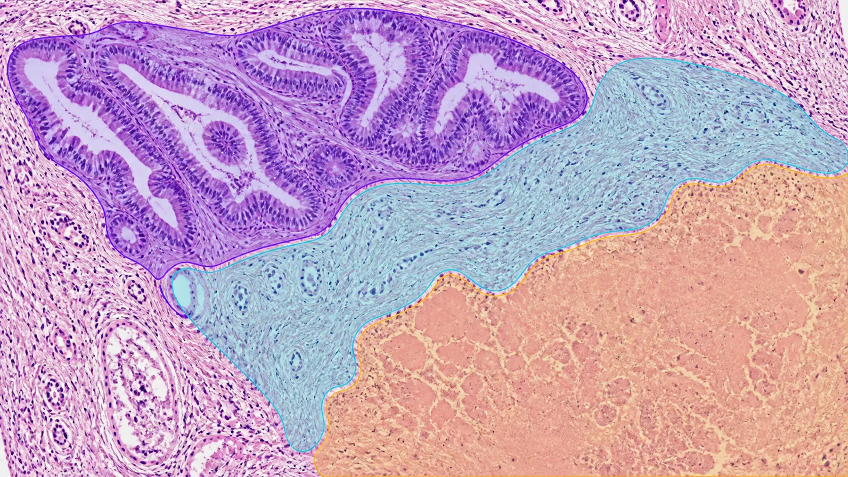
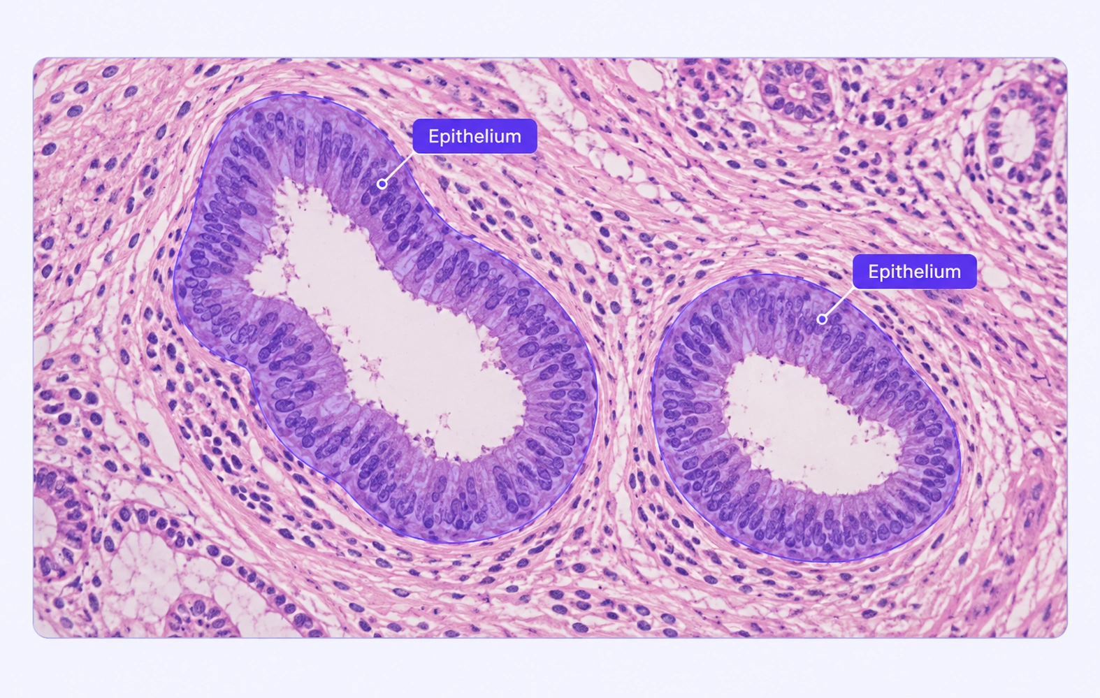
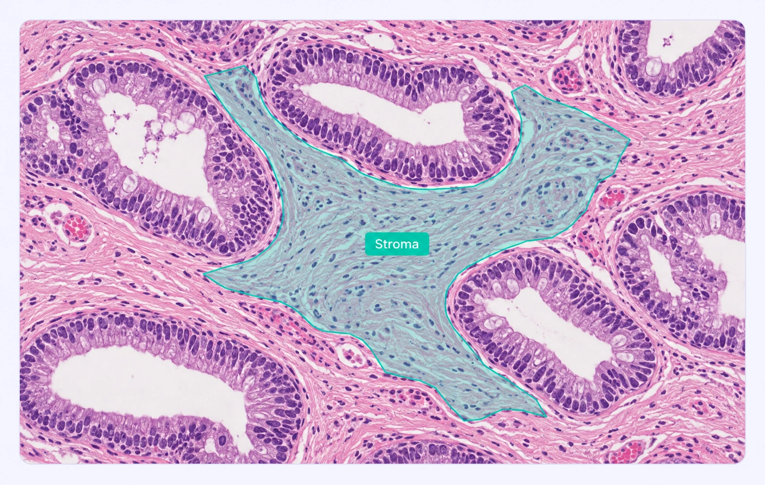
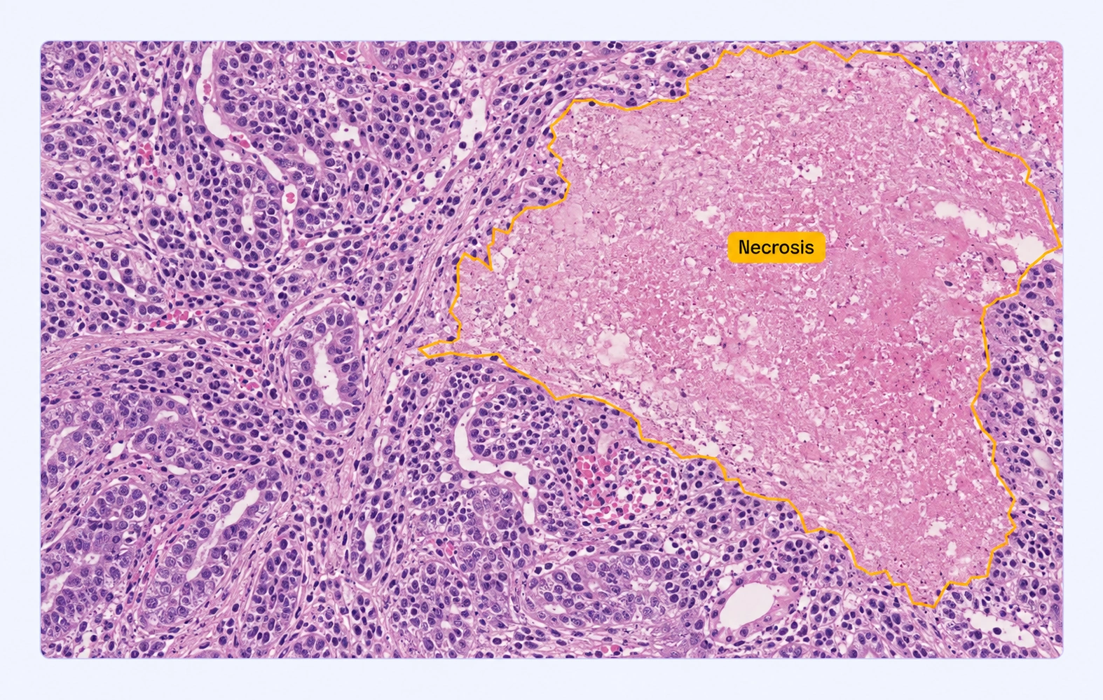
Label the area belonging to each tissue class with segmentation regions in the original slide coordinates.
Outline morphological regions with polygons when the training task requires explicit boundaries.
It labels regions of tissue in histology images according to a defined class scheme, creating training examples for computational pathology segmentation models.
Yes. Define the tissue types, compartments, and properties that match your protocol rather than relying on a fixed list of labels.
Yes. Tiled navigation lets annotators move between broader tissue context and detailed regions while working in the source slide.
A mask describes a labeled area, while a contour explicitly traces its boundary. Choose the geometry that matches your model and annotation guidelines.
Use your protocol’s uncertainty rules and reviewer comments to handle ambiguous transitions. Specialist review can determine the accepted class and boundary.
Yes. Define tissue or background classes where your dataset needs a foreground distinction, and apply consistent rules around slide edges and empty regions.
Supported inputs include SVS, AVS, NDPI, SCN, BIF, SVSLIDE, tiled TIFF, and single-file OME-BigTIFF slides. Tiled navigation lets teams inspect large slides at different zoom levels.
Reviewers inspect cellular or tissue boundaries in slide context, discuss uncertain regions, and return corrections through the configured workflow. Shared classes keep the annotation protocol consistent.
Yes. Pathology labels retain the original full-resolution raster coordinate space. Supported formats include COCO, YOLO, and Unitlab Unified Export Format, depending on the annotation data you need.


We use cookies to improve your experience. See our Privacy Policy and Cookie Policy.