
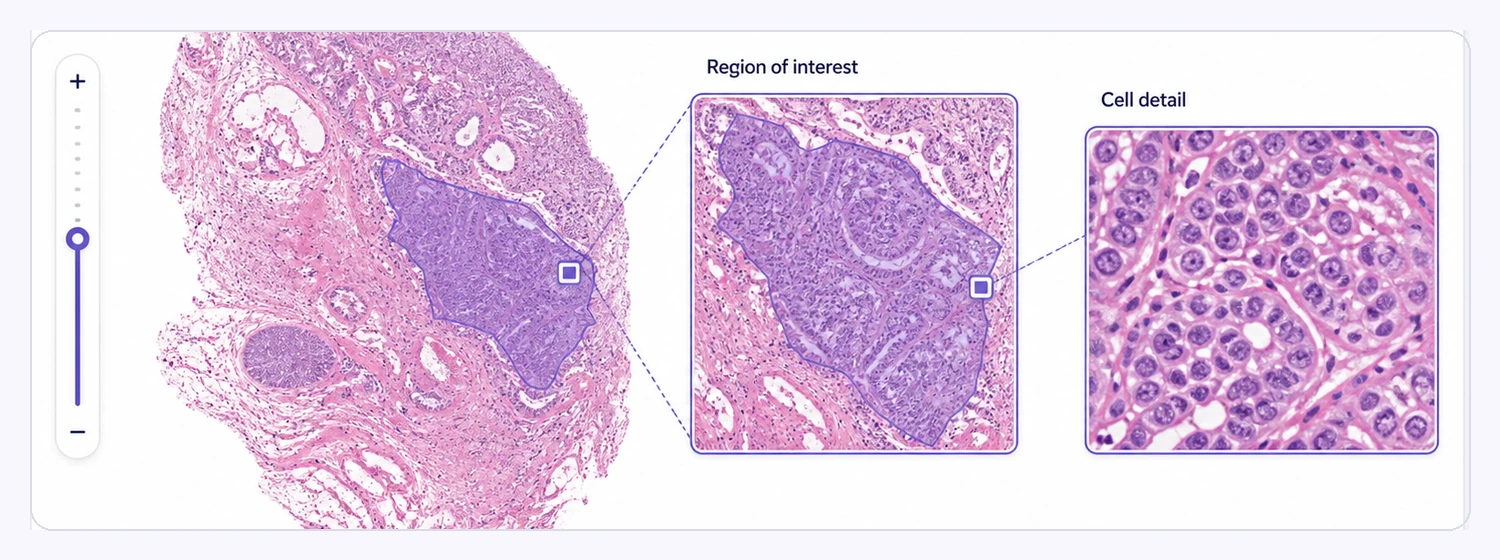
Whole-Slide Image Support
Handle gigapixel whole-slide images natively while preserving full-resolution pathology data.
Whole-slide imagesMulti-resolution zoomRegion navigation

Annotate whole-slide images (WSI) for histopathology, tissue regions, cells, nuclei, biomarkers, and digital pathology AI with precise segmentation and expert review workflows.
Work with whole-slide pathology images, move from tissue overview to cellular detail, organize labels with structured ontologies, and accelerate precise annotation with AI-assisted tools.
Handle gigapixel whole-slide images natively while preserving full-resolution pathology data.
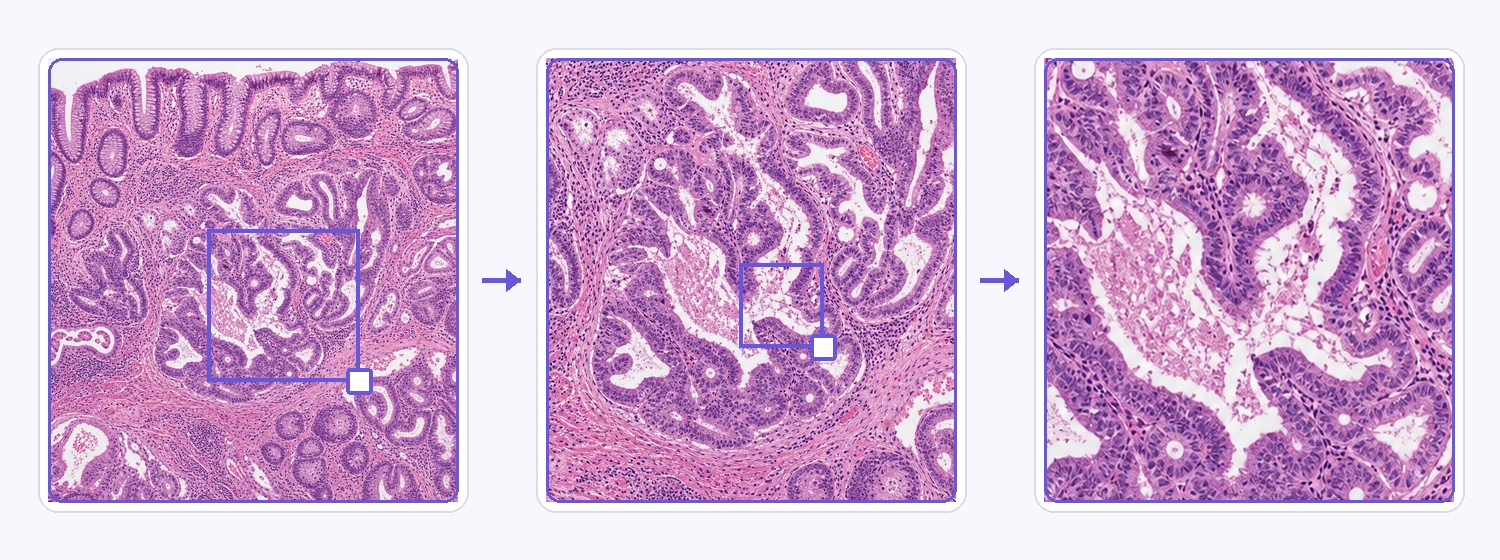
Navigate seamlessly from whole-tissue overview to cellular-level detail across multiple resolution levels.
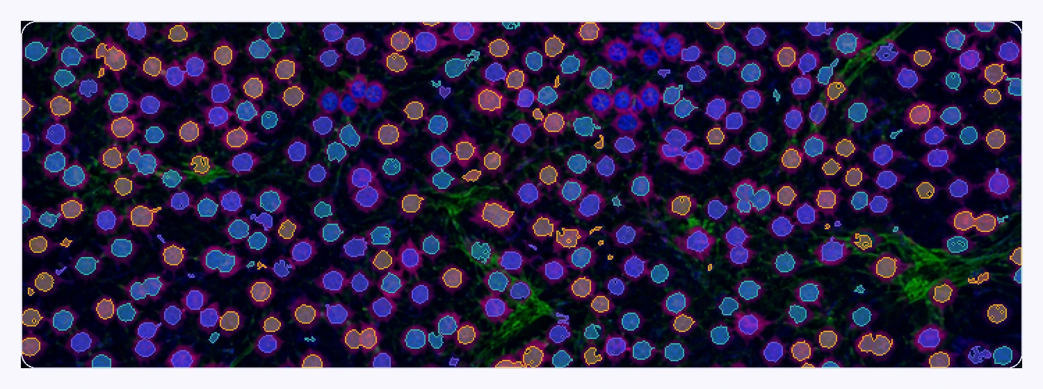
Create, render, and manage thousands of cells, nuclei, regions, and other annotations across massive pathology slides.
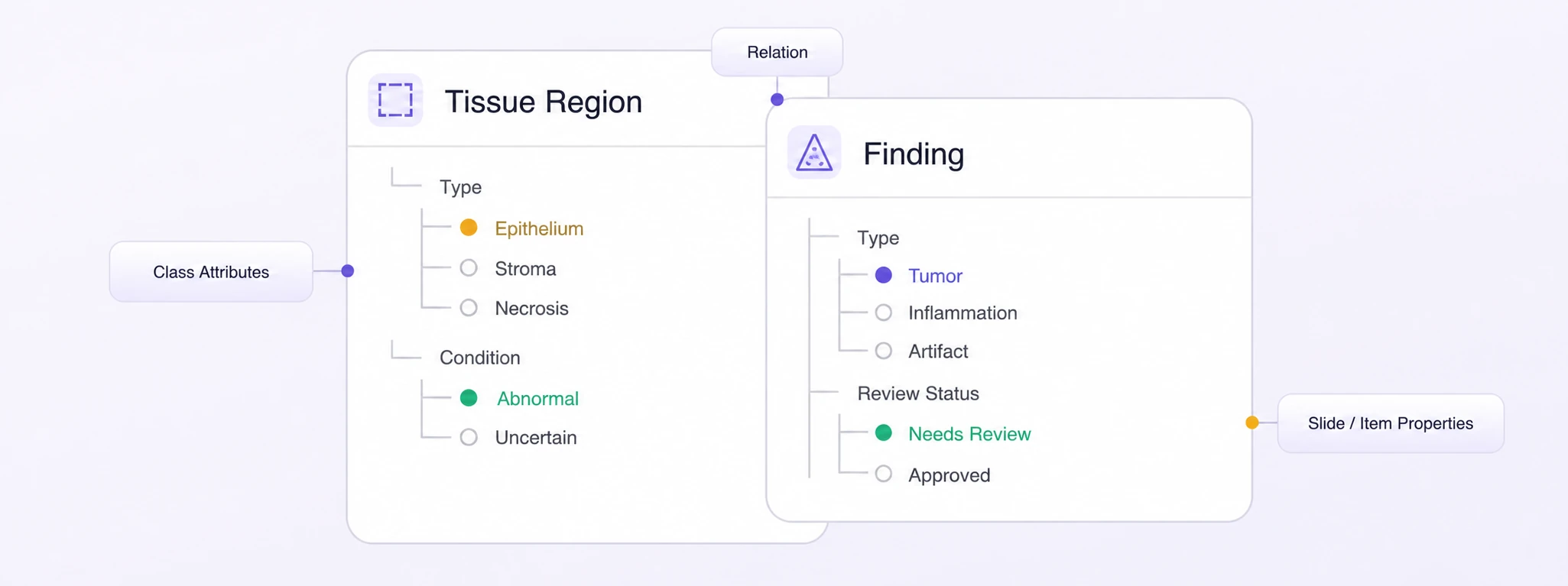
Define nested pathology classes, attributes, relations, and slide-level properties for consistent annotation workflows.
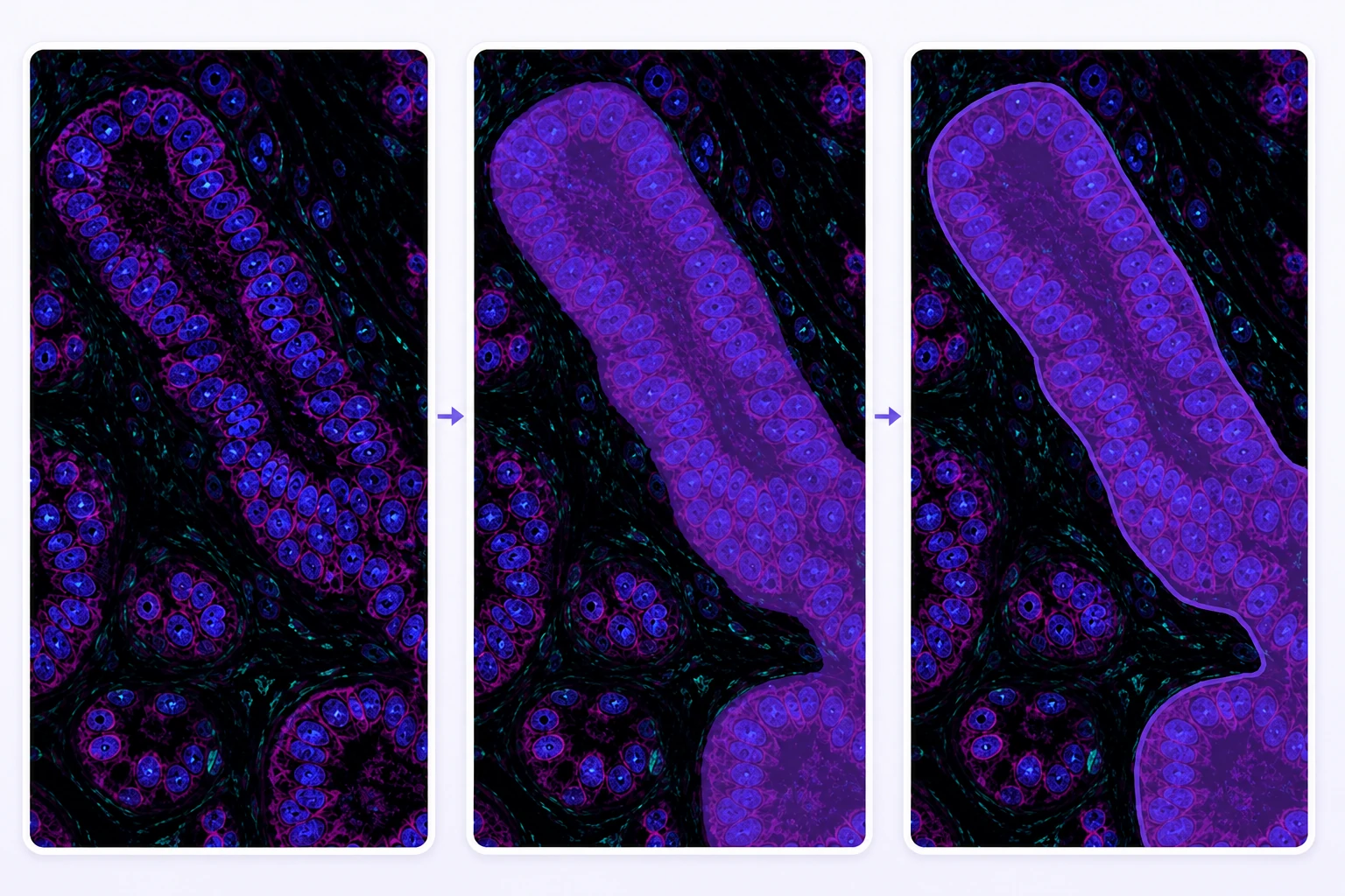
Precisely delineate cells, nuclei, tumors, tissue regions, and complex microscopic structures at pixel-level accuracy.
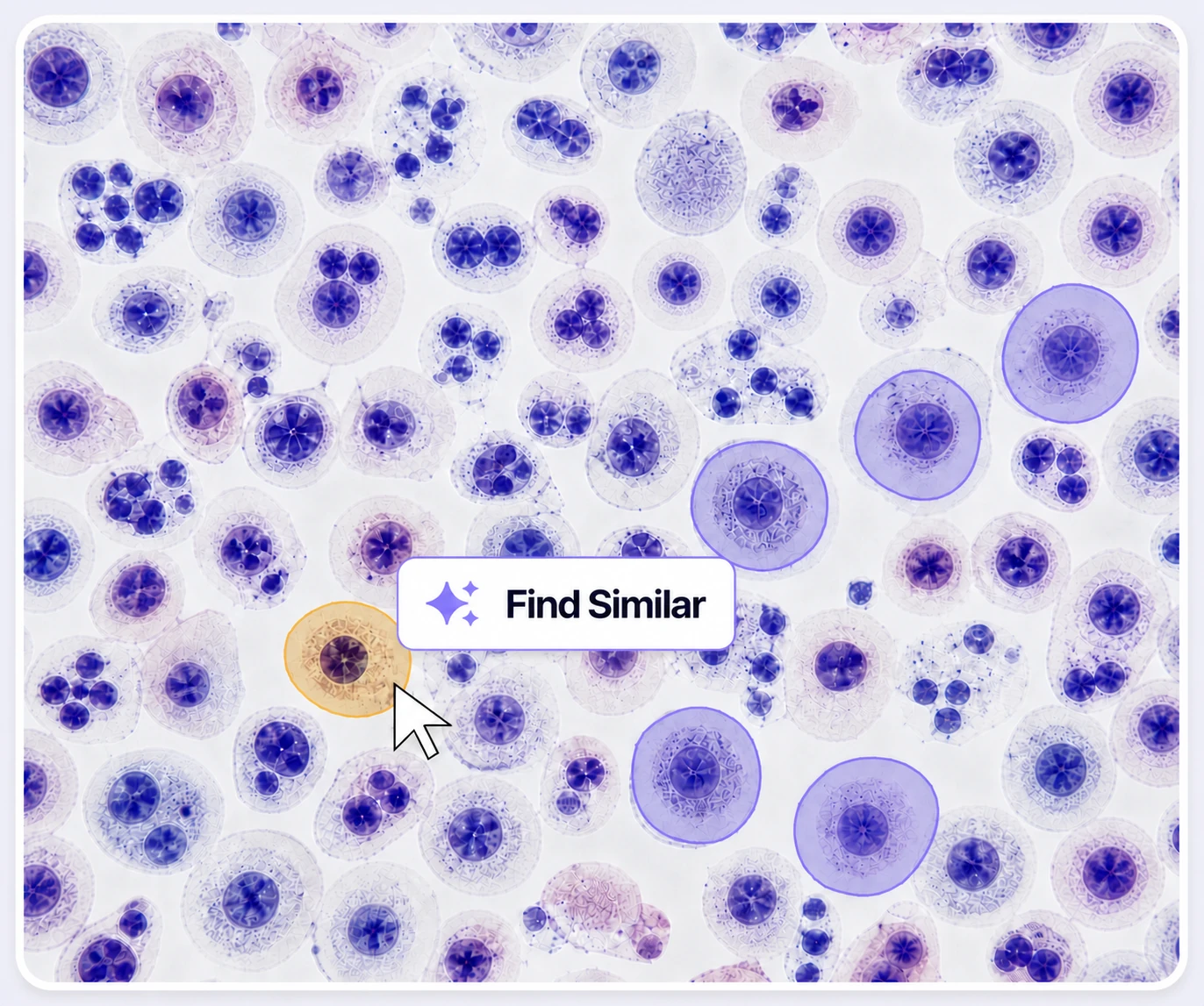
Accelerate pathology annotation with Find Similar, Magic Touch, and automated labeling tools for repetitive structures.
Annotate complex pathology datasets faster with AI-assisted automation, scalable workflows, and lower operational costs.
Batch, Find Similar, Prompt Auto-Labeling, and automated workflows reduce repetitive manual pathology labeling.
On average, 90% of pathology labels are pre-labeled automatically, then reviewed and refined by humans.
The cost per accepted label can be up to 5× lower as curation, annotation, and QA are automated.
Create regions of interest, tissue segmentation masks, polygons, cell and nuclei instances, point markers, classifications, slide properties, and relations for pathology workflows.
Mark rectangular regions of interest for tissue, tumor, lesion, cell cluster, or review areas.
Capture pixel-level tissue, tumor, lesion, necrosis, or biomarker regions with editable masks.
Outline irregular tissue regions, tumor margins, glands, and other structures with editable polygons.
Label individual cells and nuclei as distinct instances for detection, counting, and morphology analysis.
Trace tissue boundaries, margins, vessels, and elongated structures with connected vertices.
Mark exact cell centers, nuclei, glands, and other microscopic landmarks on a slide.
Assign slide-, region-, or finding-level classes such as tissue type, grade, stain, or biomarker status.
Label properties that describe the complete slide or item, including source, quality, cohort, and project-specific attributes.
Connect findings and tissue regions to capture project-defined contextual relationships.
Compare labels on the same tissue regions, check them against approved references, and resolve issues at the right magnification. Keep expert decisions connected to the original slide evidence.
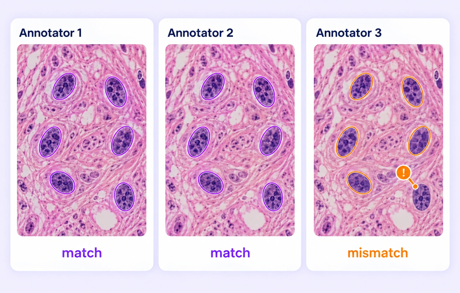
Compare independent labels on the same histology region. Identify omitted nuclei, differing tissue boundaries, and class disagreements for expert review.
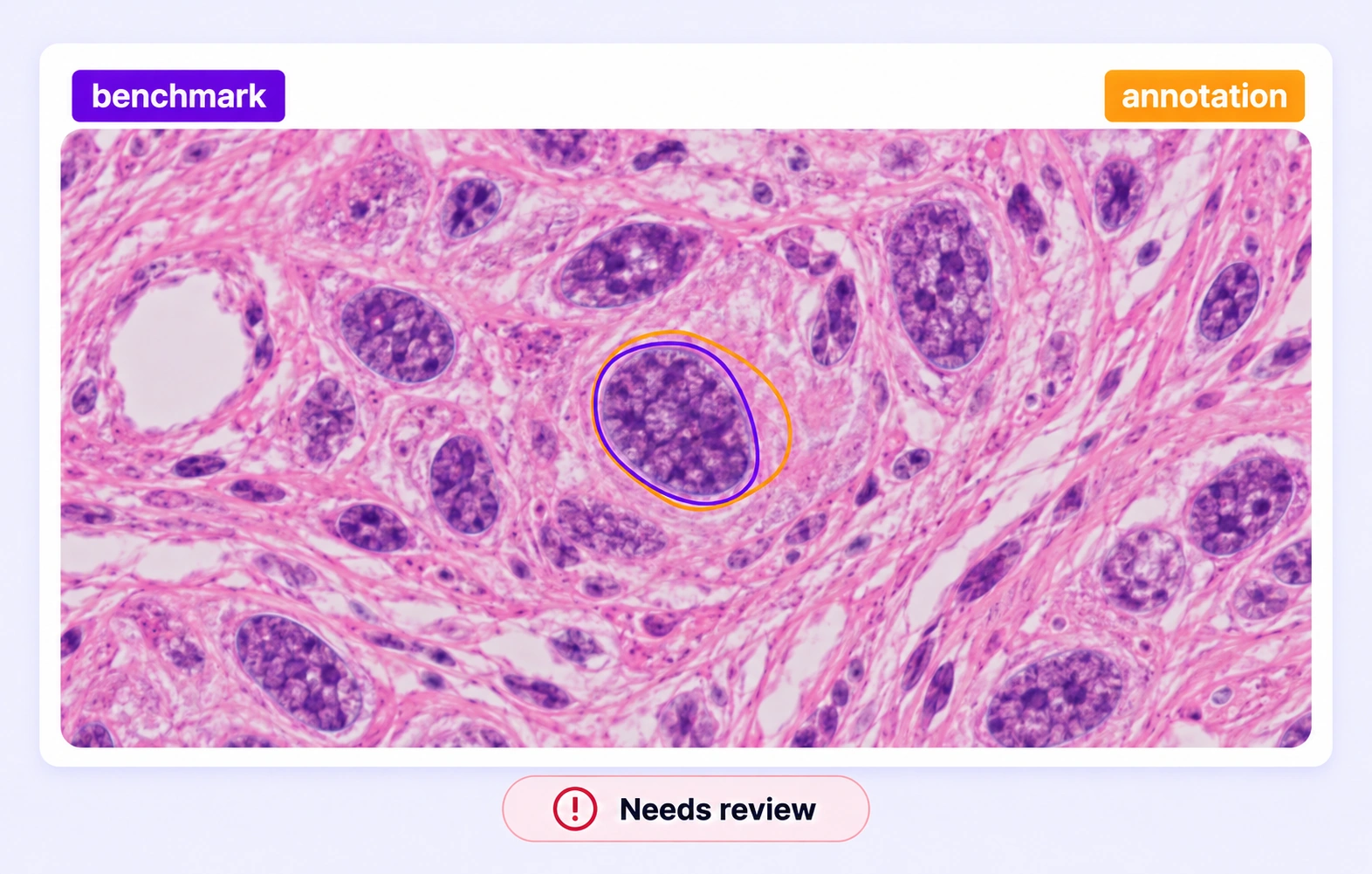
Check pathology annotations against approved references hidden from annotators. Evaluate supported boundaries and classes using the project's quality criteria.
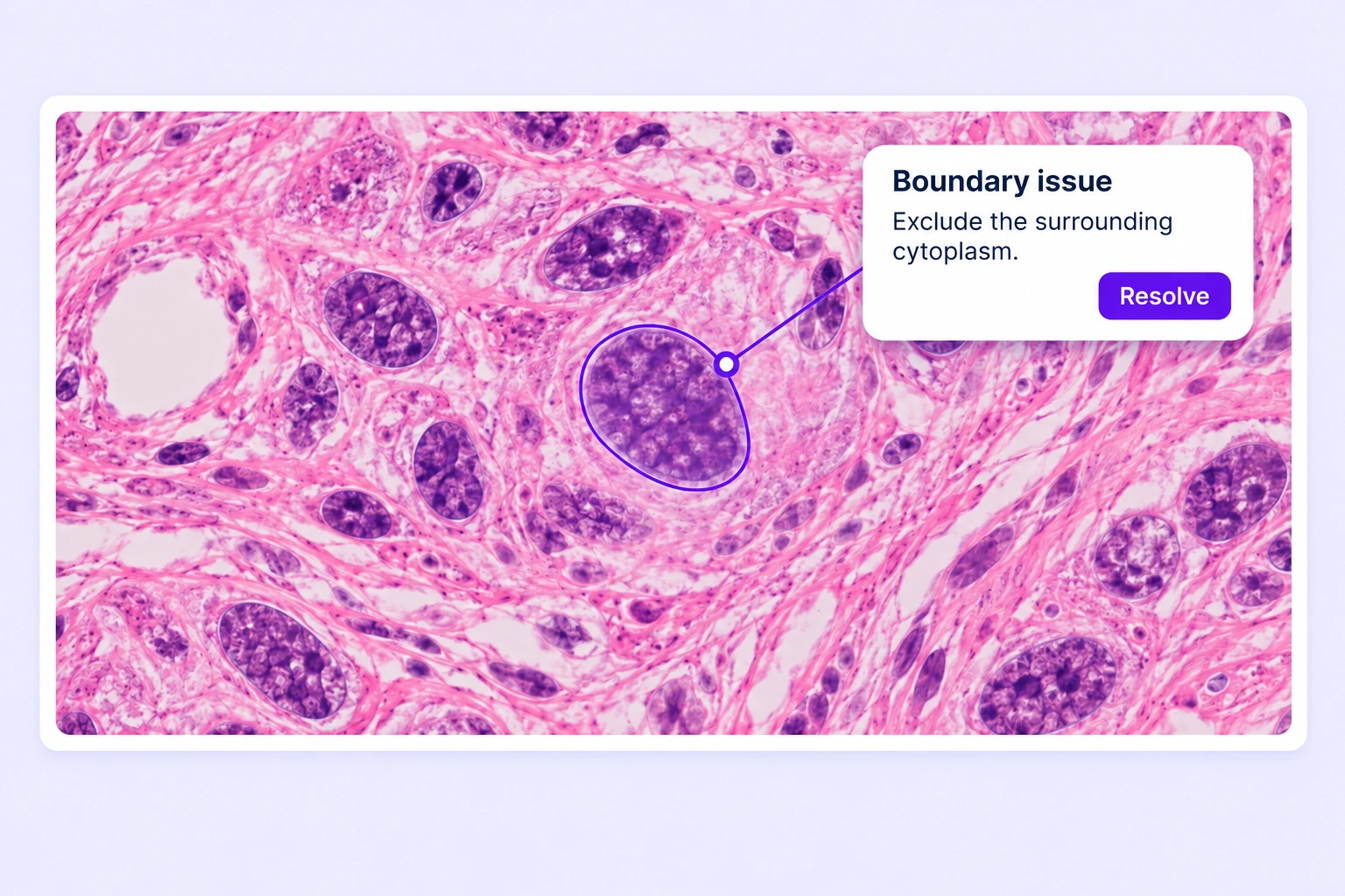
Review missing properties and disputed nucleus or tissue boundaries in slide context. Correct the affected annotation and resolve feedback before approval.
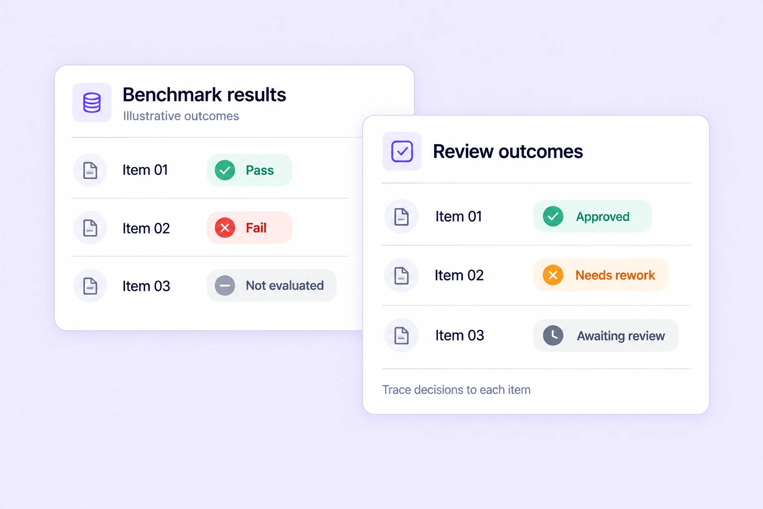
Track benchmark results, consensus outcomes, and review decisions across slide annotation tasks. Identify recurring issues and focus expert review on difficult regions.
Search, version, and inspect pathology datasets, connect AI models, and move annotations through review and approval.
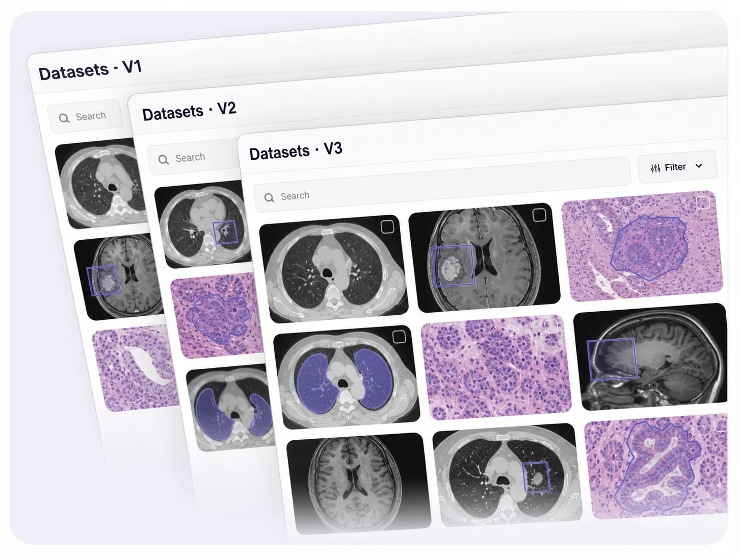
Create and manage dataset versions as pathology data evolves. Track changes, assign work, and keep every version auditable and production-ready.
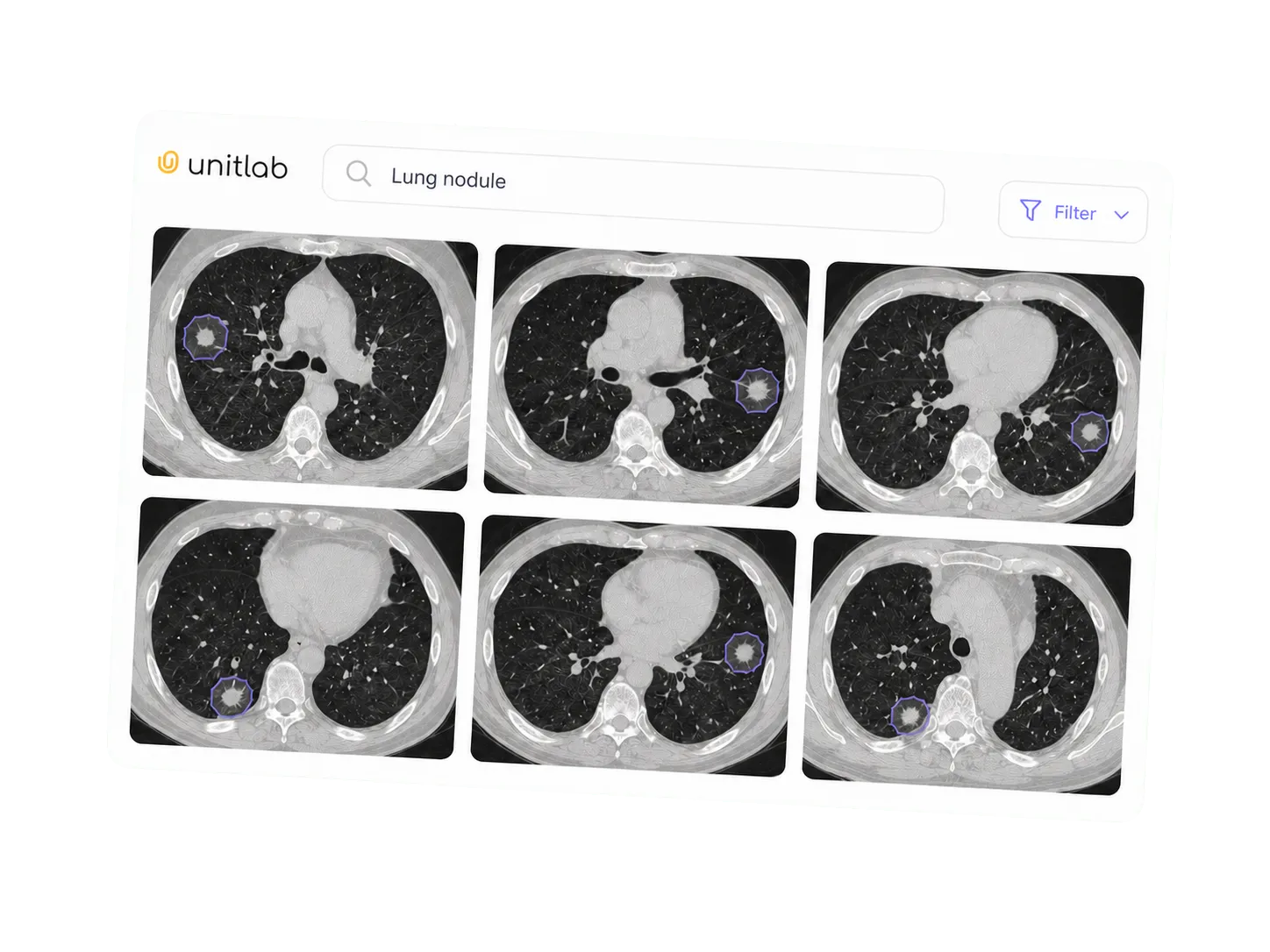
Explore large pathology datasets through semantic understanding instead of manual filters. Find relevant cases and slide regions across defined conditions.
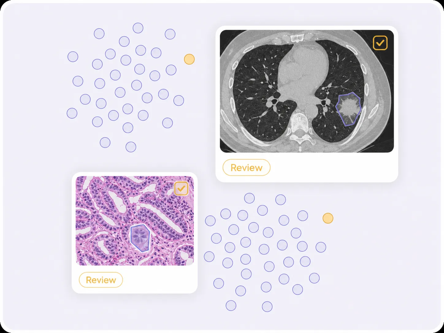
Visualize dataset structure, identify outliers and labeling issues, and improve pathology-data quality before training.
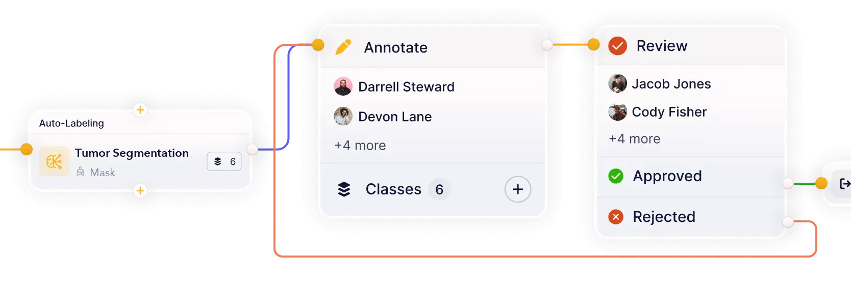
Build workflows that connect models, annotation, review, and quality assurance in one continuous loop. Reduce handoffs and keep datasets moving from labeling to approval.
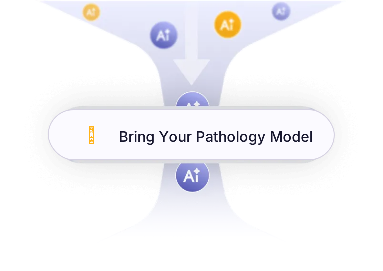
Connect your own AI models for pre-labeling and model-assisted annotation. Improve accuracy and iterate faster on real-world pathology datasets.
Build high-quality whole-slide and cellular datasets for tissue analysis, tumor modeling, and biomarker research.
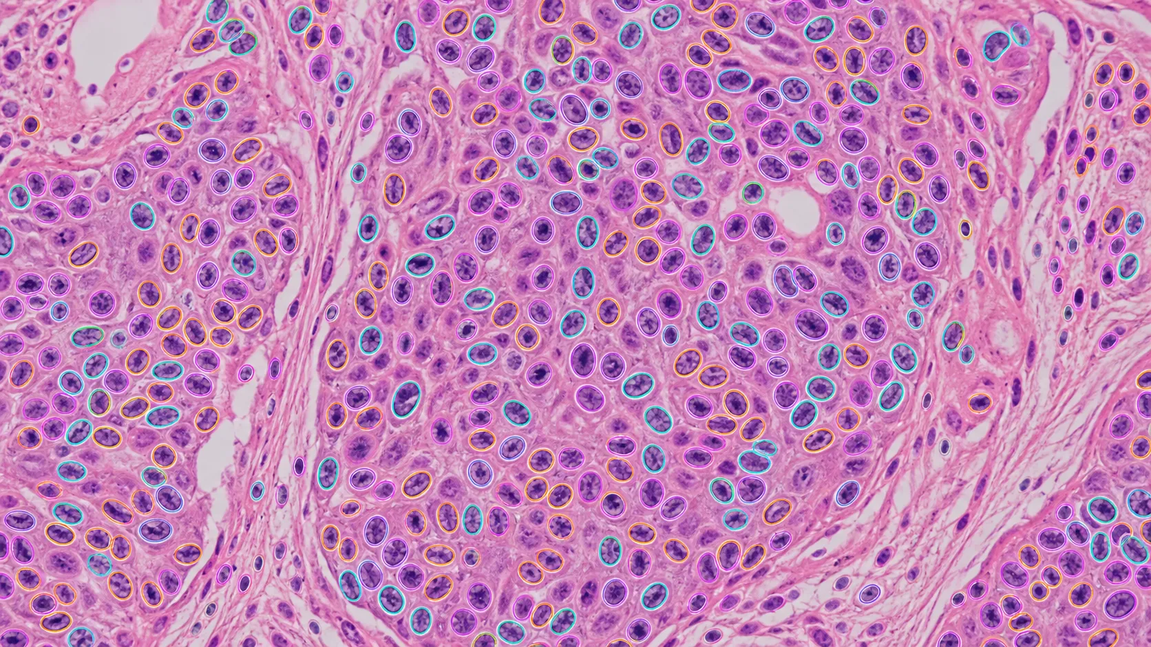
Annotate individual cells, nuclei, and cellular structures across whole-slide pathology images for detection and quantification.
Explore cell & nuclei analysis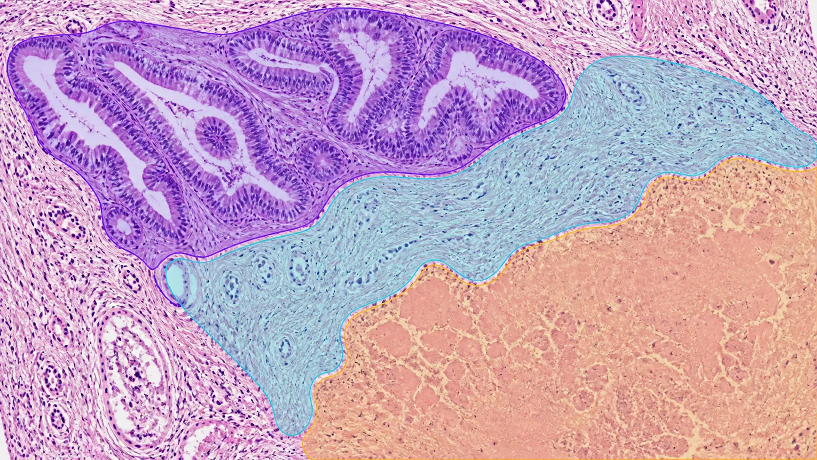
Label tissue types, compartments, and morphological regions across gigapixel histology slides.
Explore tissue segmentation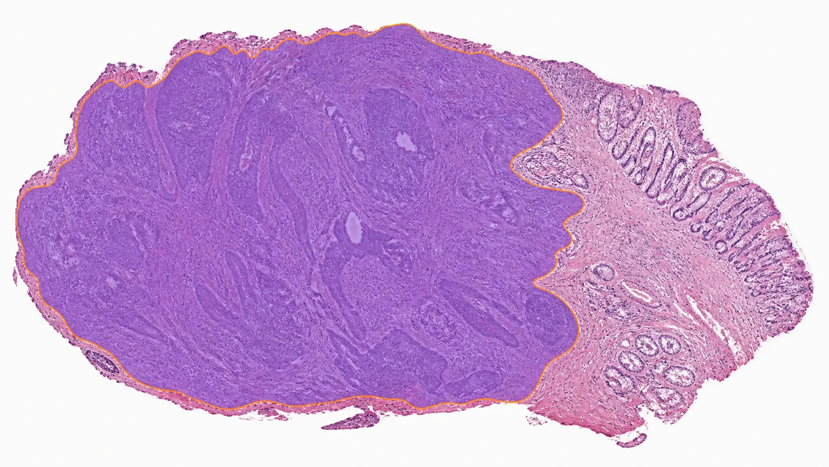
Define tumor boundaries, margins, necrotic regions, and surrounding tissue for computational pathology models.
Explore tumor region identification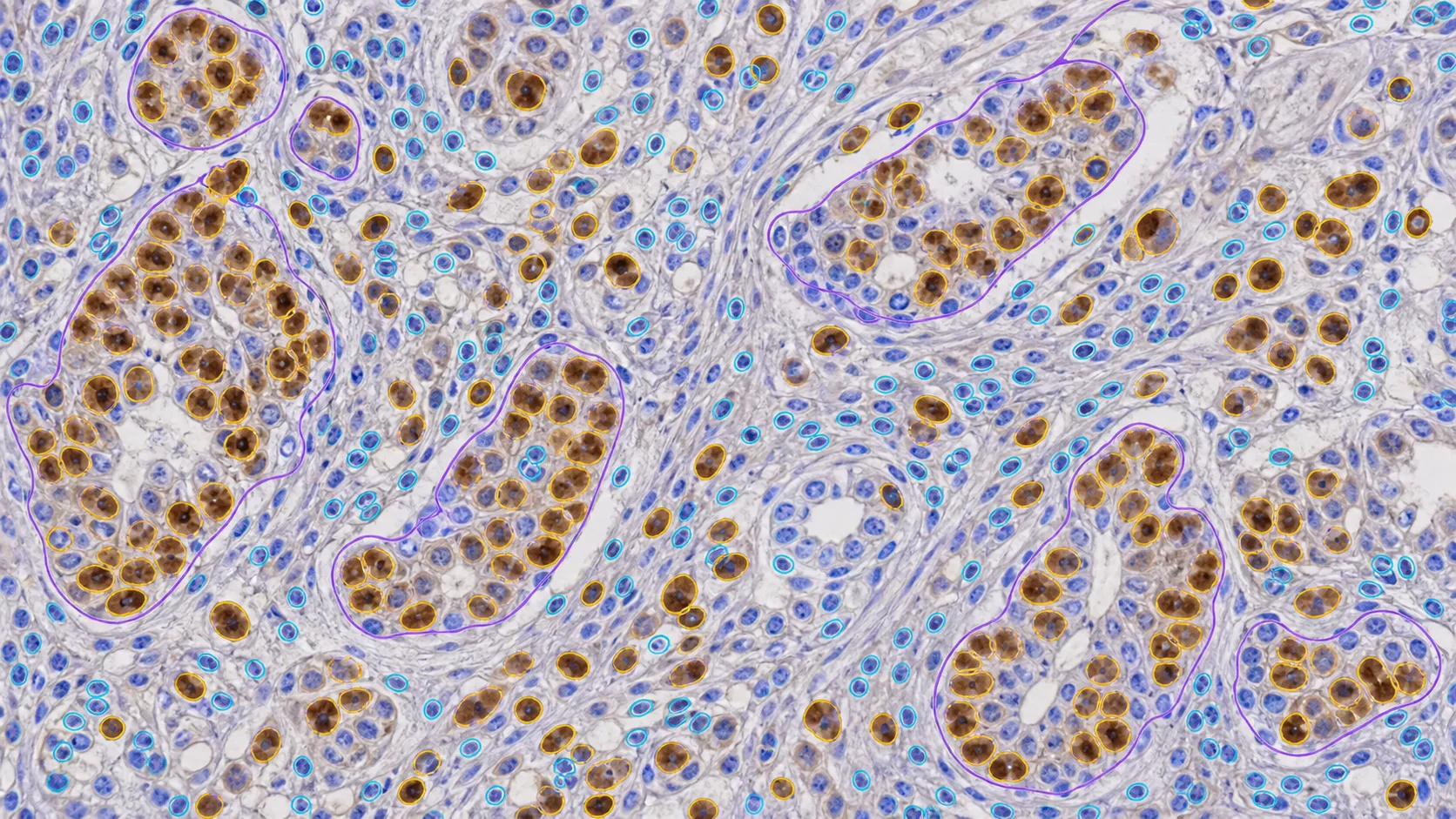
Label positive and negative staining, cellular regions, and expert-defined staining categories for IHC training datasets.
Explore IHC & staining annotationAnswers about WSI and histopathology annotation, cells, nuclei, tissue segmentation, digital pathology ontologies, quality workflows, custom models, and dataset operations in Unitlab.
Talk with the Unitlab teamUnitlab supports ROI boxes, tissue segmentation masks, polygons, cell and nuclei instances, point markers, boundaries, classifications, slide properties, and relations. Teams define labels and rules in reusable ontologies for consistent whole-slide image annotation.
Pathology annotation documentationUse a built-in or integrated image model to generate a segmentation or polygon prediction, then review and refine the boundary before the annotation moves through quality review.
Auto-annotation documentationReviewers can inspect the full image and focused regions, compare labels against the ontology, leave issues and comments, and use annotation history to track what was reviewed or corrected.
Annotation review documentationNested ontologies define classes, classifications, properties, and relations. Class properties describe annotated regions, while slide or item properties capture context that applies to the complete image.
Properties and relations documentationConfigurable workflows route pathology tasks through annotation, review, rework, and approval. Instructions, assignments, comments, issues, annotation history, dataset versions, and releases keep quality decisions traceable for individual experts and enterprise teams.
Annotation and review documentationYes. Unitlab can bring custom models into the annotation workflow to generate pre-labels and predictions. Annotators review and correct model output instead of starting from zero, while human approval remains part of the governed quality process.
Model integration documentationYes. Teams can curate pathology data with metadata filters, semantic search, embeddings, similarity, and outlier discovery, then annotate selected samples, review results, and publish controlled dataset versions for reproducible AI development.
Dataset management documentationAnnotate, review, and manage complex pathology image data in one AI-assisted workspace. Move from raw images to governed datasets with model-assisted labeling and built-in quality control.


We use cookies to improve your experience. See our Privacy Policy and Cookie Policy.