
Built-in auto-labeling tools and automated workflows reduce manual annotation work.
Automate curation, dataset management, and versioning so engineers can focus on models.
Curated data, reusable ontologies, and quality control reduce annotation rework.
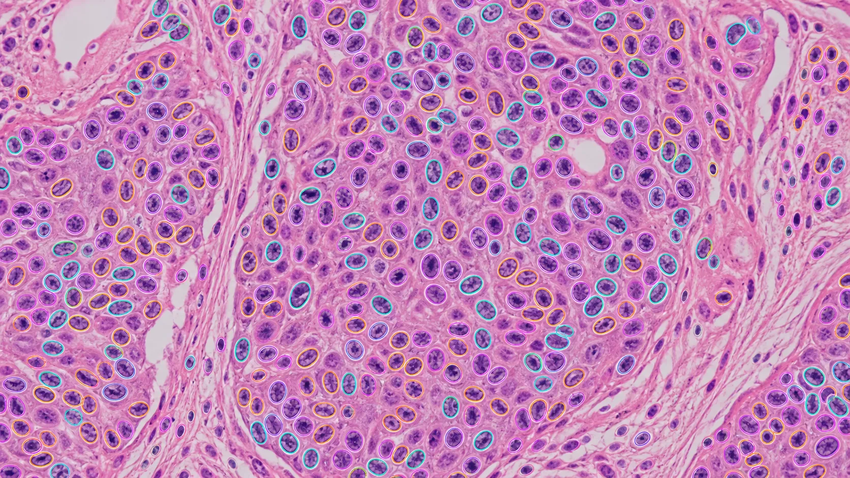
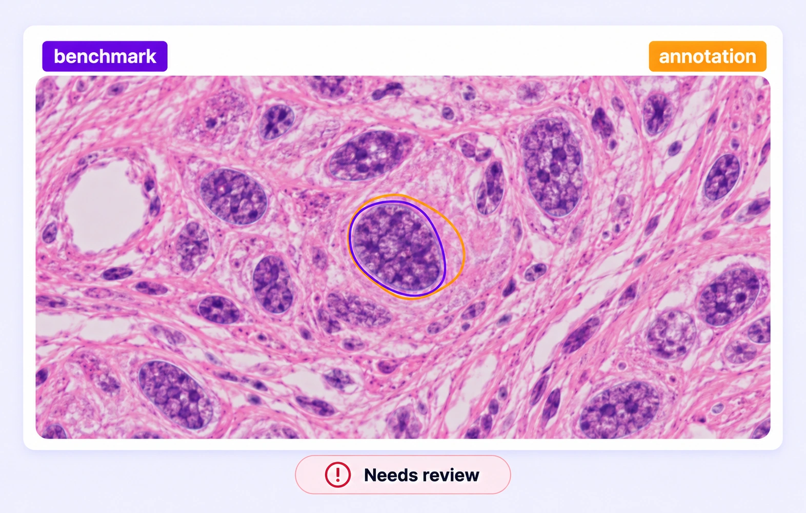
Check whether each in-scope nucleus has a separate, structure-aligned annotation. Resolve omitted instances, merged neighbors, and contours extending into surrounding cytoplasm.
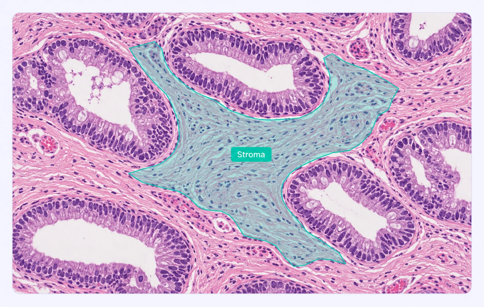
Review epithelial, stromal, and other protocol-defined tissue regions in the original slide. Refine boundaries around glands and adjacent compartments while preserving the surrounding context.
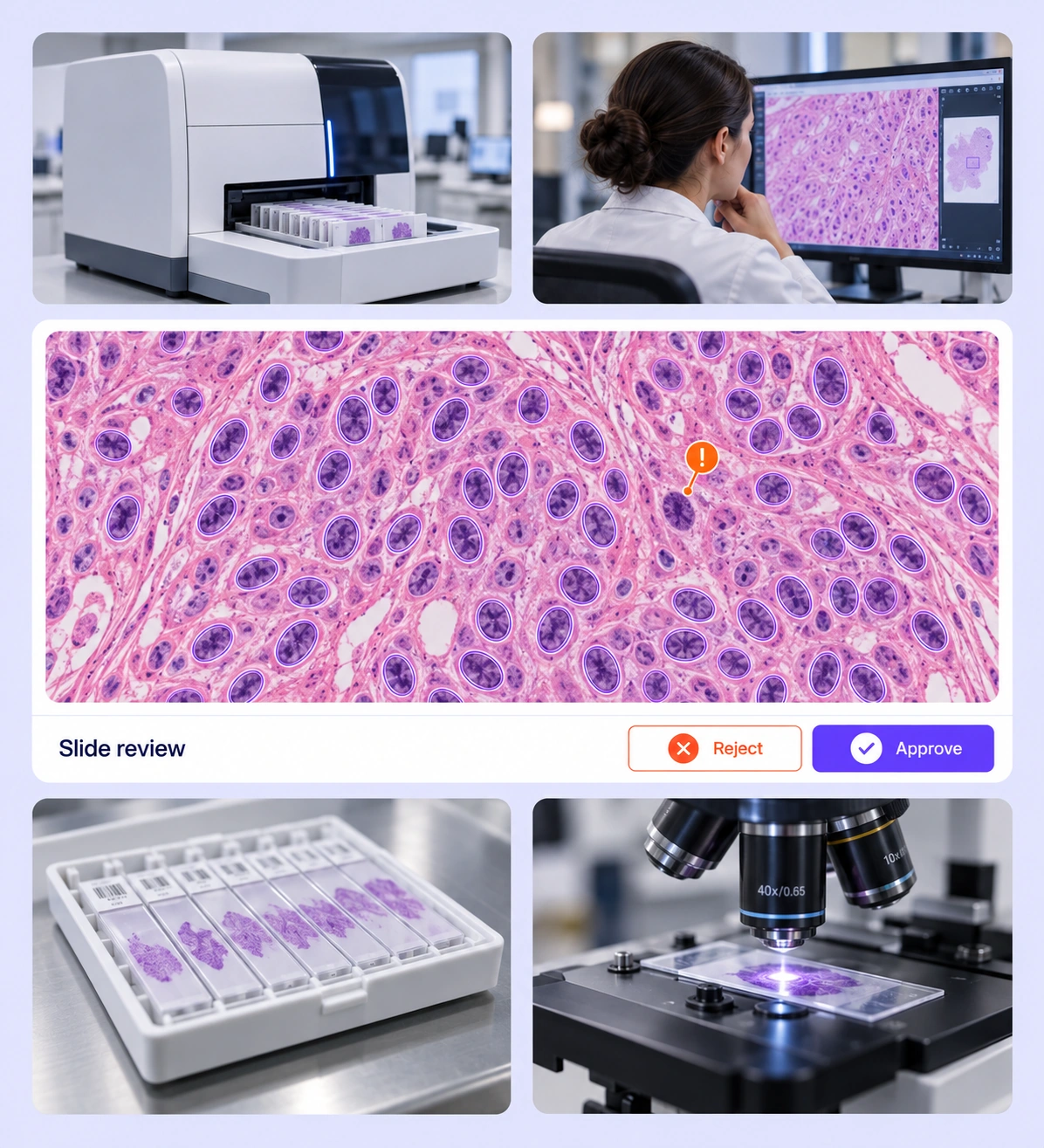
It is the review of whole-slide and histology labels for completeness, consistent class definitions, and alignment with visible structures. Unitlab supports the annotation and review workflow; qualified reviewers define and apply the study protocol. QA overview
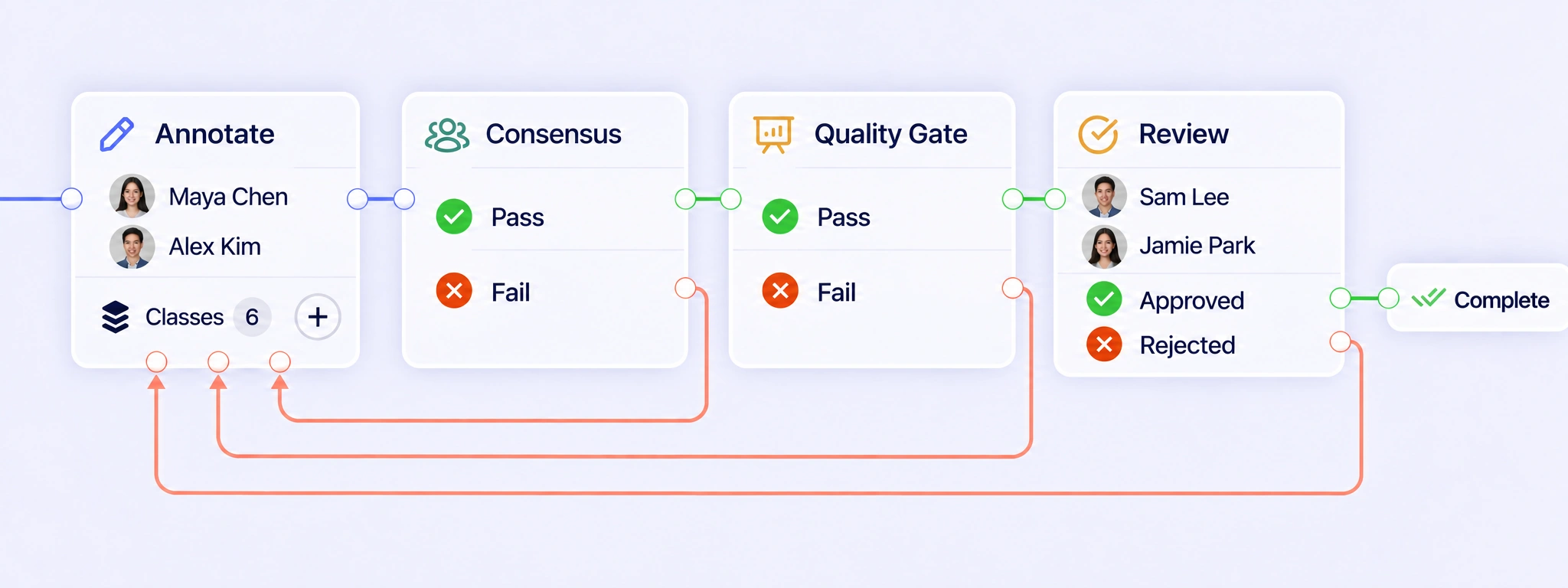
Configure annotation, Consensus, Quality Gate, expert review, and completion around the study protocol. Failed checks or rejected slide annotations can return for correction through a rework path. Review the revised labels in their original tissue context before the item proceeds through the required checks again. QA workflows
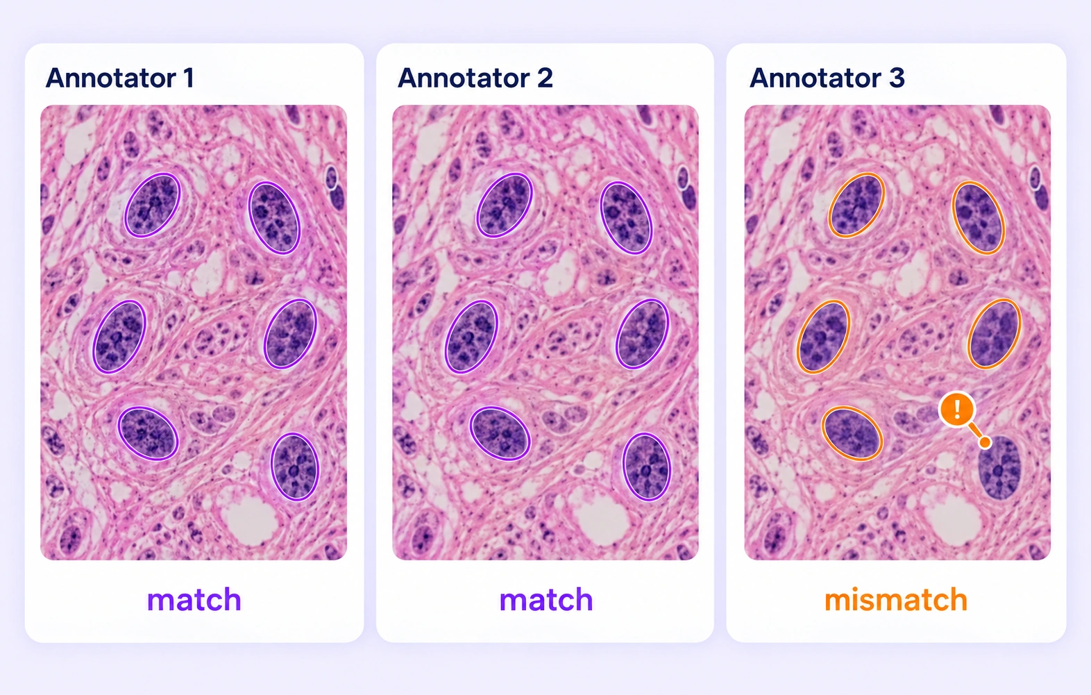
Consensus compares independent submissions of the same pathology input under the configured settings. Differences can reveal omitted nuclei, inconsistent classes, or tissue boundaries that need review. An expert inspects the microscopic evidence and applies the study protocol; agreement alone does not establish the correct interpretation. Consensus guide
Quality Gate compares eligible submissions with an approved, frozen answer key for the same pathology item. The reference remains hidden from annotators and reflects the study’s accepted labeling rules. Configure Pass, Fail, and Not evaluated routes, including expert review when the available comparison data is incompatible or incomplete. Quality Gate guide
Teams can inspect supported tissue regions, cell or nucleus instances, point markers, and slide or annotation properties. The study protocol should define the target structures, class meanings, and inclusion rules. Reviewers check both whether each expected structure is labeled and whether its geometry matches the visible evidence. Review stages
Yes. The pathology workbench supports large whole-slide imagery with zoom and multi-resolution viewing. Reviewers can inspect a local cell or boundary, then consider the surrounding tissue. Use the viewing scale required by the study so local detail and broader context inform the labeling decision. Review stages
Define how the study distinguishes individual instances and treats partially visible or overlapping nuclei. Reviewers can inspect local boundaries, look for merged instances or missed nuclei, and correct the affected labels. Apply the same inclusion and separation rules across the dataset rather than relying on annotator preference. Review stages
Have qualified reviewers inspect the disputed region in its surrounding tissue context and apply the study’s class definitions. Record a clear convention for similar examples, including how uncertainty should be represented through supported properties. These decisions establish consistent training labels without treating annotation agreement as a diagnosis. Review stages
Create a reviewed dataset release and select an export supported by the pathology input and annotation geometry. Check that tissue and cell classes, instance rules, and required properties match the intended experiment. Preserve the study protocol and review documentation alongside the dataset used for training or evaluation. Review stages


We use cookies to improve your experience. See our Privacy Policy and Cookie Policy.