
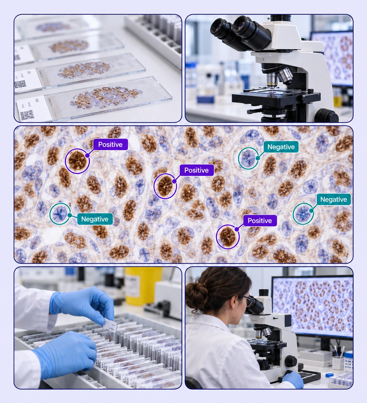
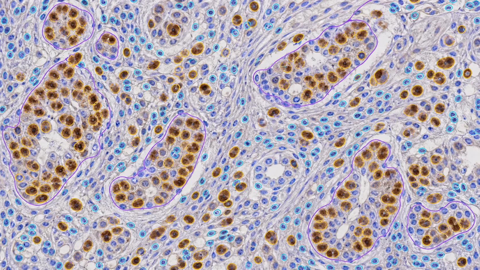
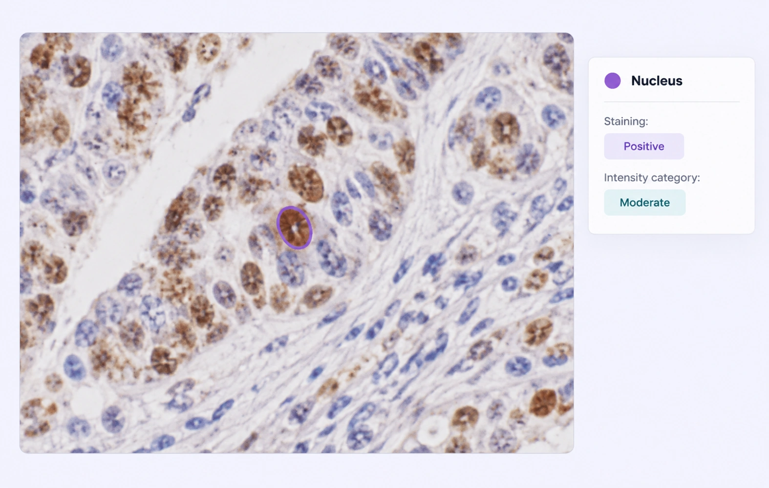
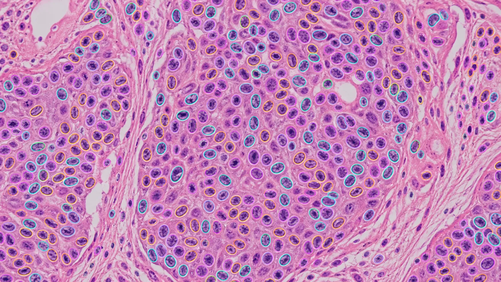
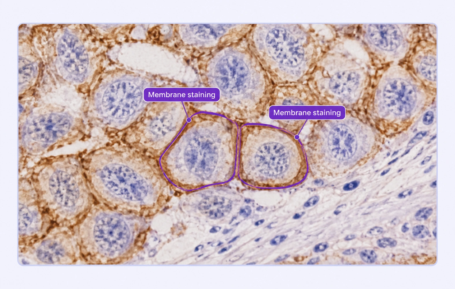
Label staining patterns and tissue regions with shared classes and expert review.
Automate curation, management, and versioning of IHC datasets.
Reusable ontologies and governed quality control reduce labeling and review rework.
Mark the cellular or tissue regions that provide the evidence for your biomarker analysis task.
Capture task-defined staining categories as structured properties attached to the relevant annotation.
It labels cellular or tissue regions and their staining categories in immunohistochemistry images, creating training data based on the interpretation protocol defined by your specialists.
Yes. Define positive, negative, or other task-specific classes with explicit visual criteria, then apply them consistently to the source images.
Yes. Use structured properties for the categories your annotation protocol defines. These are expert labels rather than automatic intensity measurements.
Yes. Reviewed regions and cellular labels can provide input to your own counting, measurement, or scoring pipeline.
Annotation records expert-defined regions and categories. Your downstream analysis pipeline can use those labels with its own validated measurement or scoring methods.
Use reference examples, define category boundaries, and review uncertain cells or regions against the same staining criteria before release.
Supported inputs include SVS, AVS, NDPI, SCN, BIF, SVSLIDE, tiled TIFF, and single-file OME-BigTIFF slides. Tiled navigation lets teams inspect large slides at different zoom levels.
Reviewers inspect cellular or tissue boundaries in slide context, discuss uncertain regions, and return corrections through the configured workflow. Shared classes keep the annotation protocol consistent.
Yes. Pathology labels retain the original full-resolution raster coordinate space. Supported formats include COCO, YOLO, and Unitlab Unified Export Format, depending on the annotation data you need.


We use cookies to improve your experience. See our Privacy Policy and Cookie Policy.