Medical image classification training data

Assign expert-defined labels to medical images and volumes. Build reviewed classification datasets for finding presence, anatomy categories, and image quality using structured item properties.
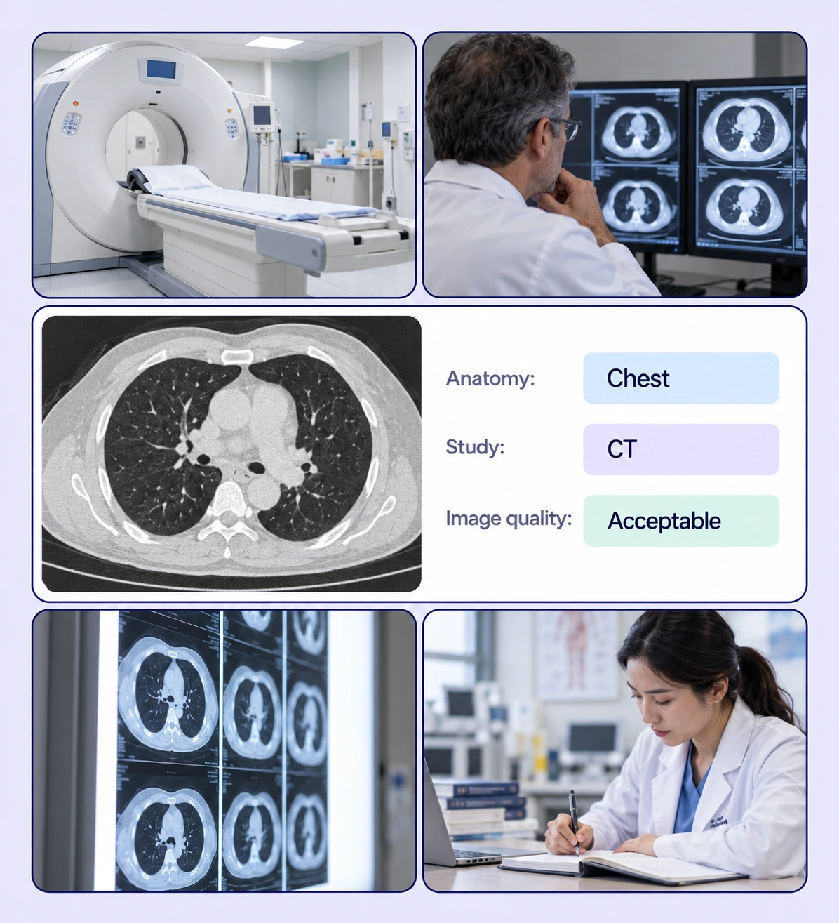
Medical Image Classification example with source-data annotations and contextual photographs.

Create clear medical image categories

Turn your classification protocol into reviewed labels for the image or volume as a whole.
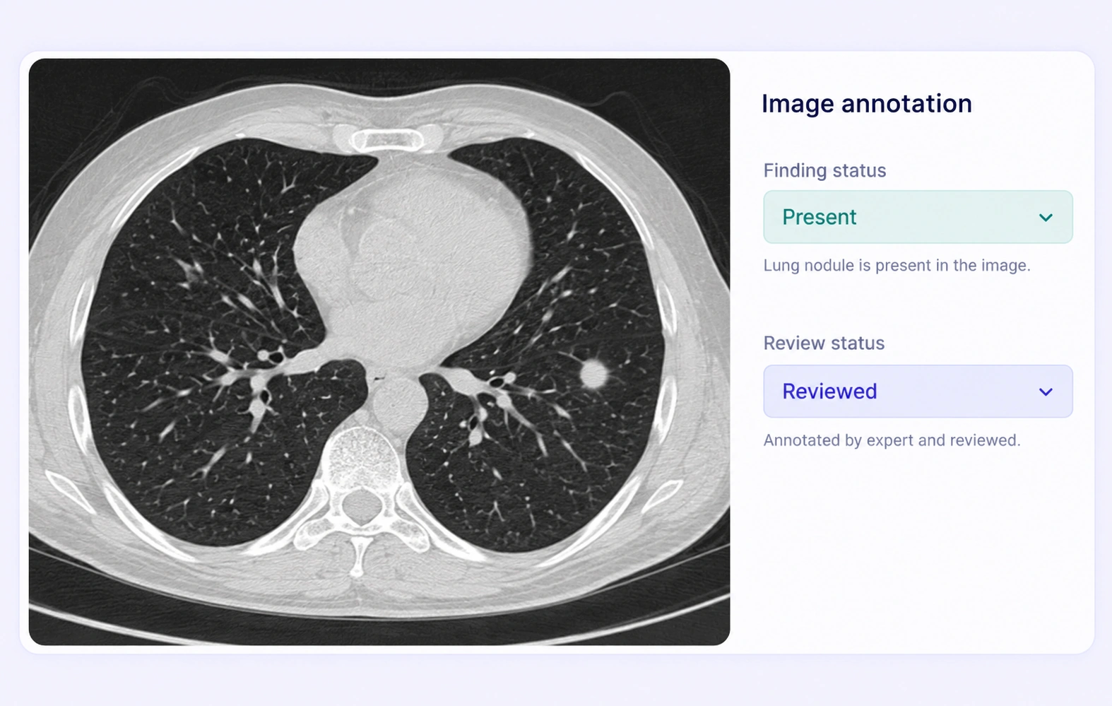
Chest CT assigned an expert-defined finding-present item label.

Finding presence labels

Record whether a task-defined finding is present, absent, or uncertain according to your expert labeling protocol.
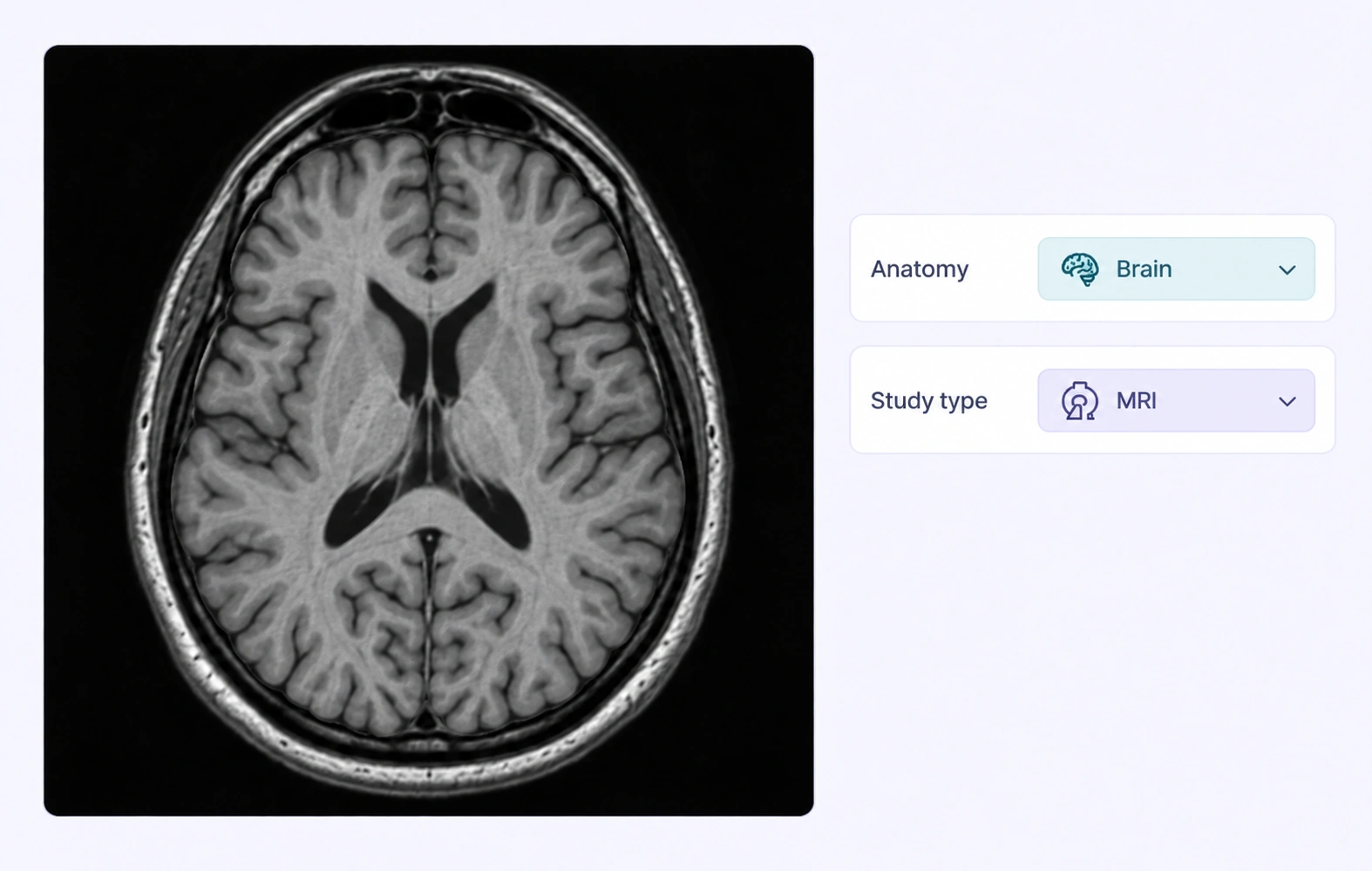
Brain MRI assigned whole-image anatomy and study-type categories.

Anatomical region categories

Classify images or volumes by the anatomical region required by your training dataset.
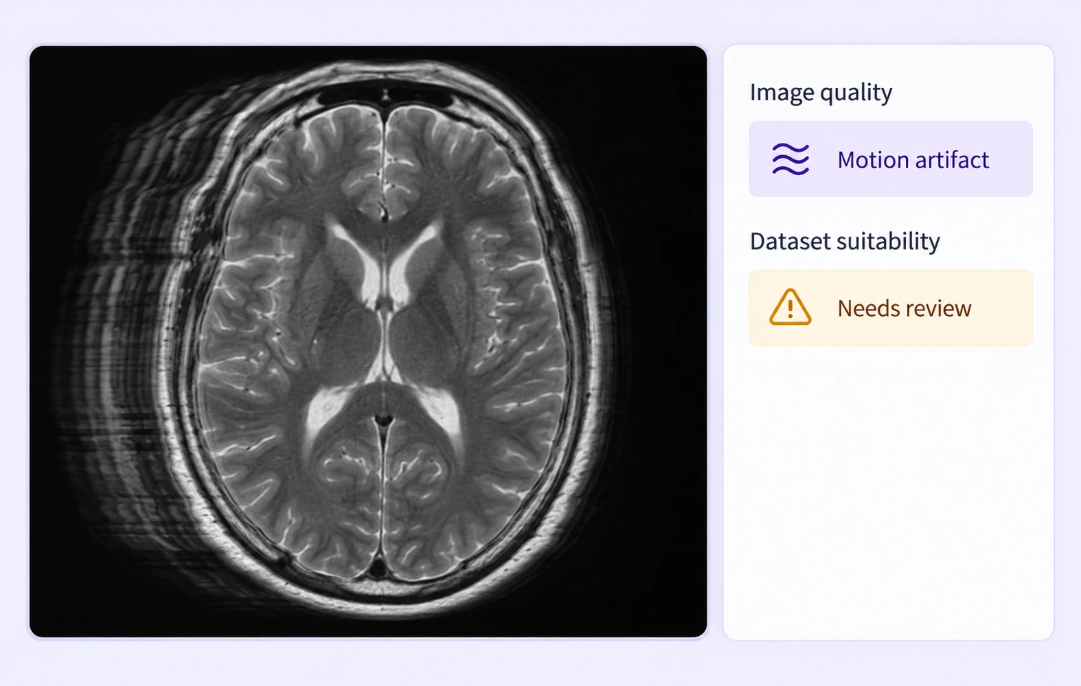
MRI example with expert-defined motion-artifact and review labels.

Image quality categories

Assign expert-defined quality categories for visible artifacts or suitability under the dataset’s inclusion criteria.
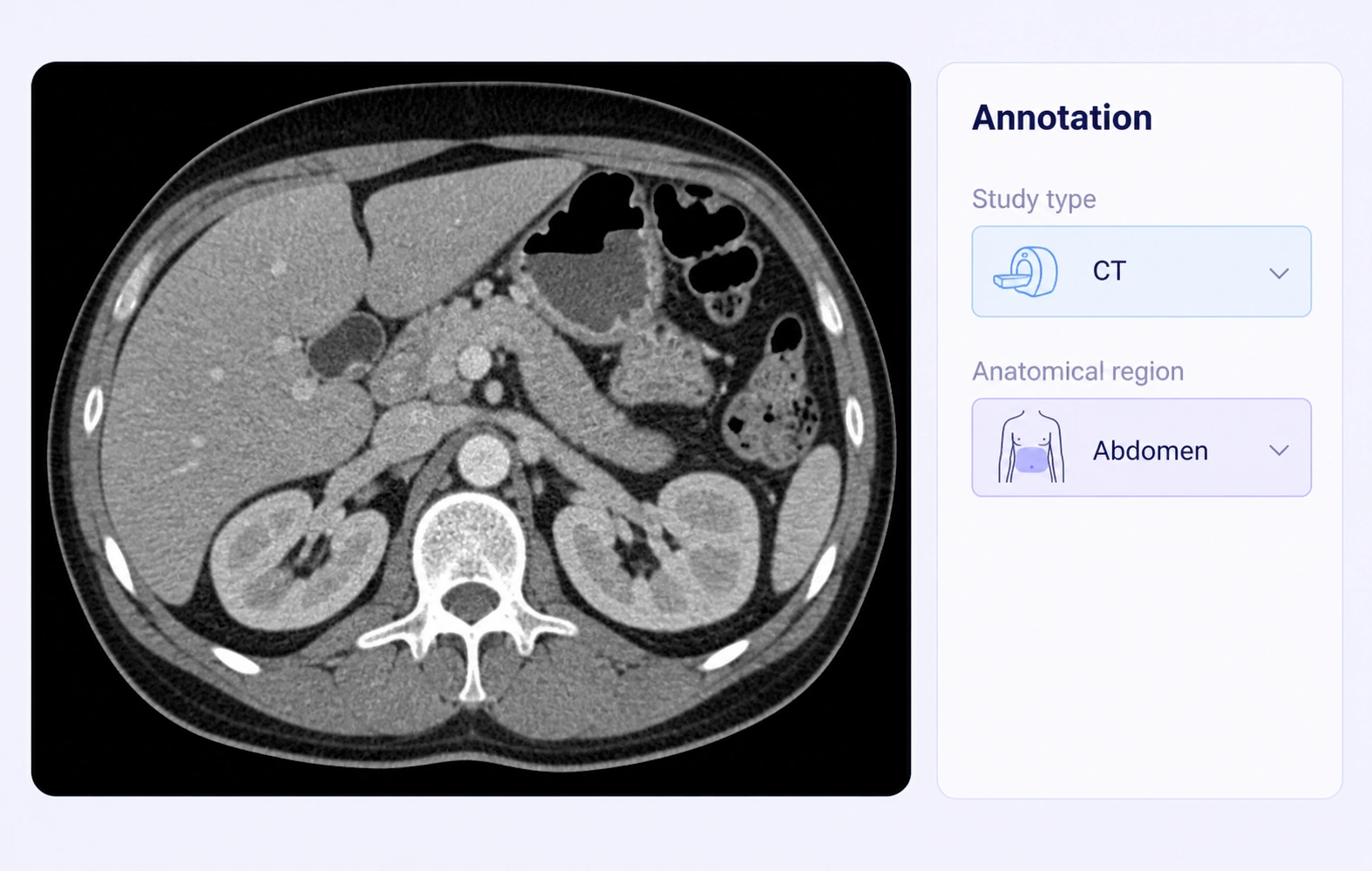
Abdominal CT with study-type and anatomical-region item labels.

Study and acquisition categories

Label task-defined study categories or acquisition characteristics as structured choices alongside the source imaging data.

Why AI Teams Choose Unitlab

Bring medical image categories, expert review, and dataset delivery into one workflow so your team can build consistent classification training data.
15X
Faster Medical Imaging Annotation
60%
Free Up AI Engineers’ Time
5X
Lower AI Development Costs

Annotation types for medical image classification

Use structured item labels for whole-image or volume categories, with optional region annotations when the task needs localized evidence.
Chest CT assigned an expert-defined finding-present item label.

Item classifications

Assign a single category or multiple allowed choices to the image or volume using the ontology’s structured properties.

Supporting regions

Add a region label when your protocol also requires the location of the evidence supporting an image-level category.
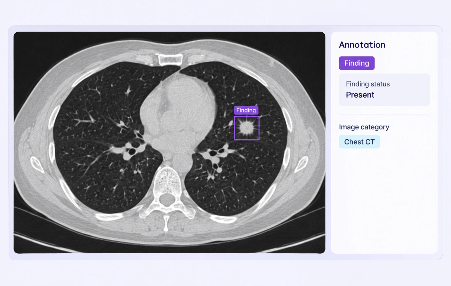
Medical image with a finding region and its supporting item classification.

Medical Image Classification FAQs

What is medical image classification annotation?

It assigns expert-defined categories to medical images or volumes so a model can learn whole-item labels such as anatomy, finding presence, or image quality.

How is classification different from segmentation?

Classification labels the image or volume as a whole. Segmentation marks the boundaries of a region within the image. A dataset can include both when required.

Can an imaging item have several labels?

Yes. Define separate properties and multi-select choices where your protocol allows more than one category to apply.

Can specialists label uncertainty?

Yes. Include an uncertainty category or another task-defined property and document how reviewers should use it consistently.

Can I include image quality labels?

Yes. Define quality categories based on visible artifacts or your dataset’s suitability criteria, then have specialists apply and review those labels.

Can I keep localized evidence with the item label?

Yes. Supported medical region annotations can accompany item properties when your protocol requires both the category and its visible location.

Which medical imaging formats are supported?

Unitlab supports DICOM, NIfTI, and NRRD inputs. DICOM slices are grouped by series into an annotation volume, with available views determined by the imaging data.

How do specialists review medical imaging labels?

Use a review stage to inspect labels against the source images, record comments, and return corrections through the workflow. Your team defines the annotation protocol and expert review criteria.

Can annotated imaging datasets be released for research?

Yes. Versioned releases and supported exports let teams deliver reviewed annotations to downstream pipelines. Select an export that preserves the geometry and metadata your research task requires.