
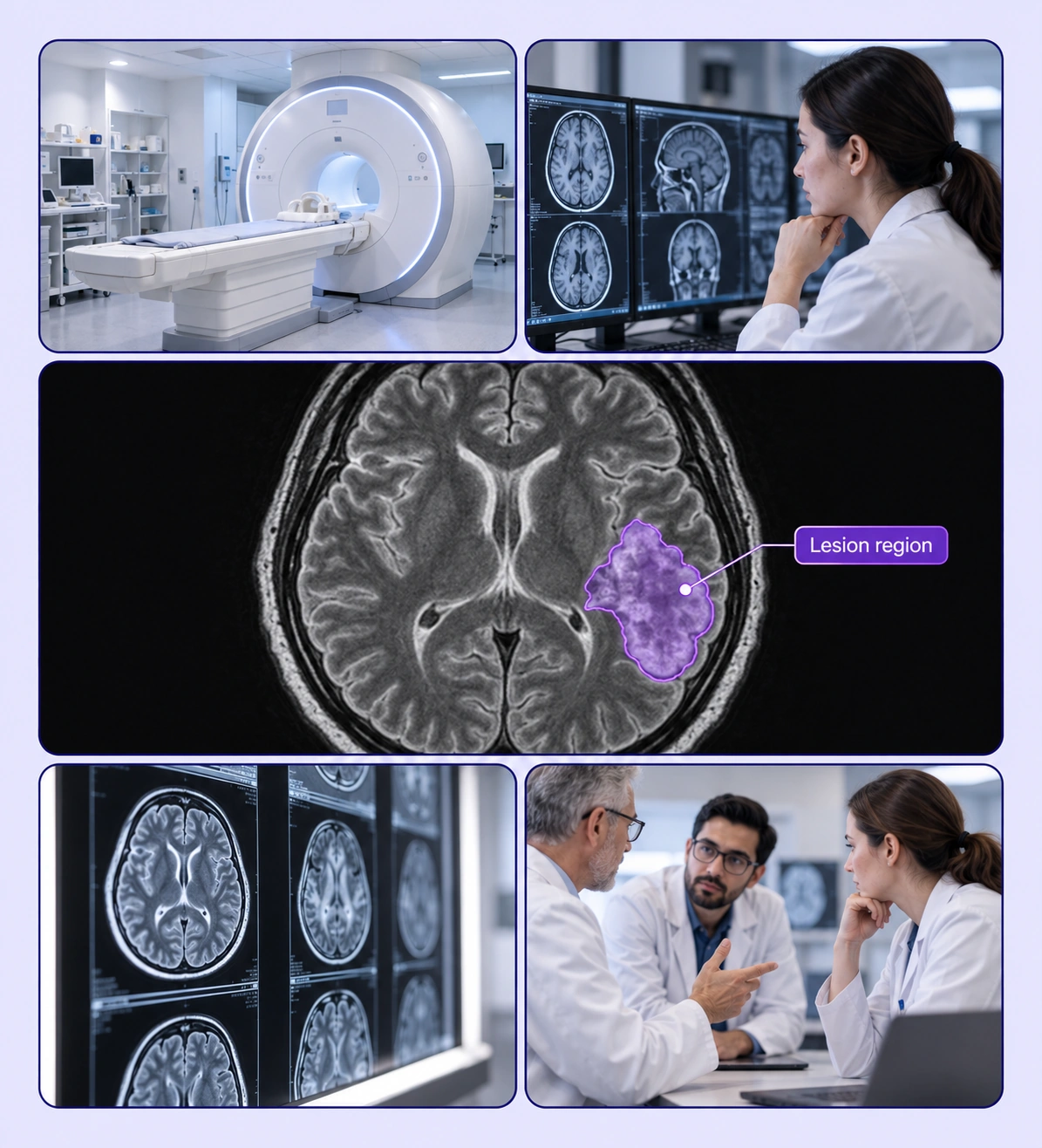
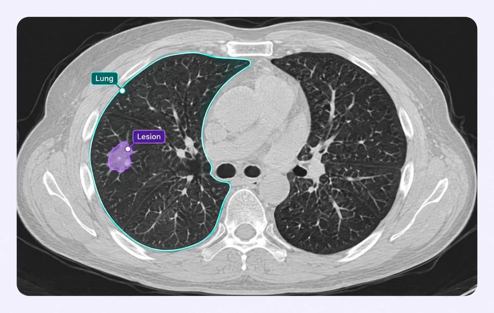
Label tumors and lesions with model-assisted tools and expert review workflows.
Automate curation, management, and versioning of lesion datasets.
Reusable ontologies and governed quality control reduce labeling and review rework.
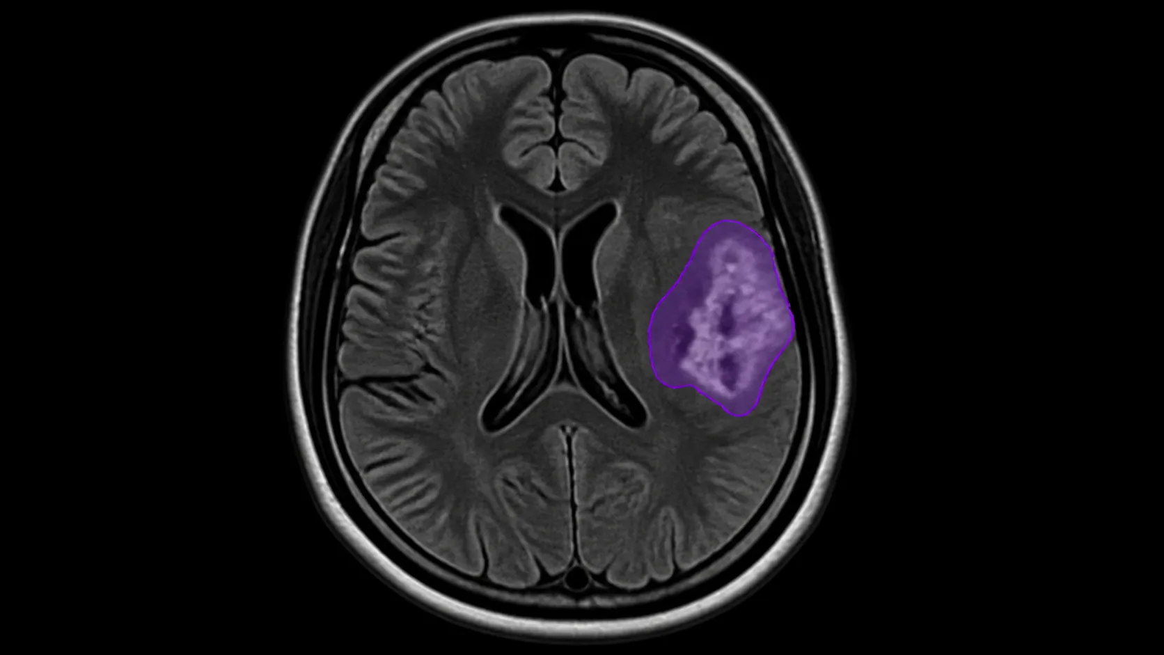
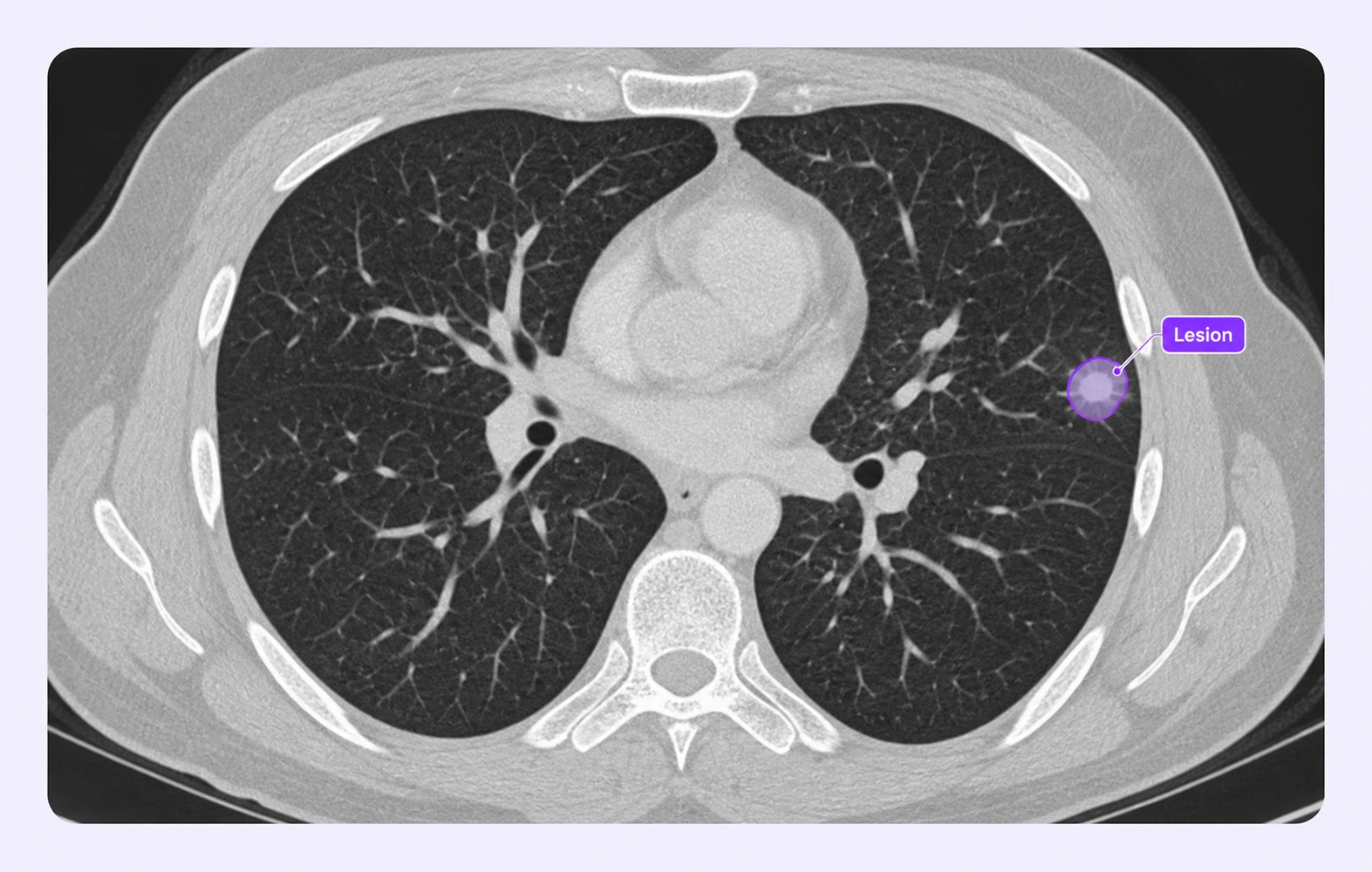
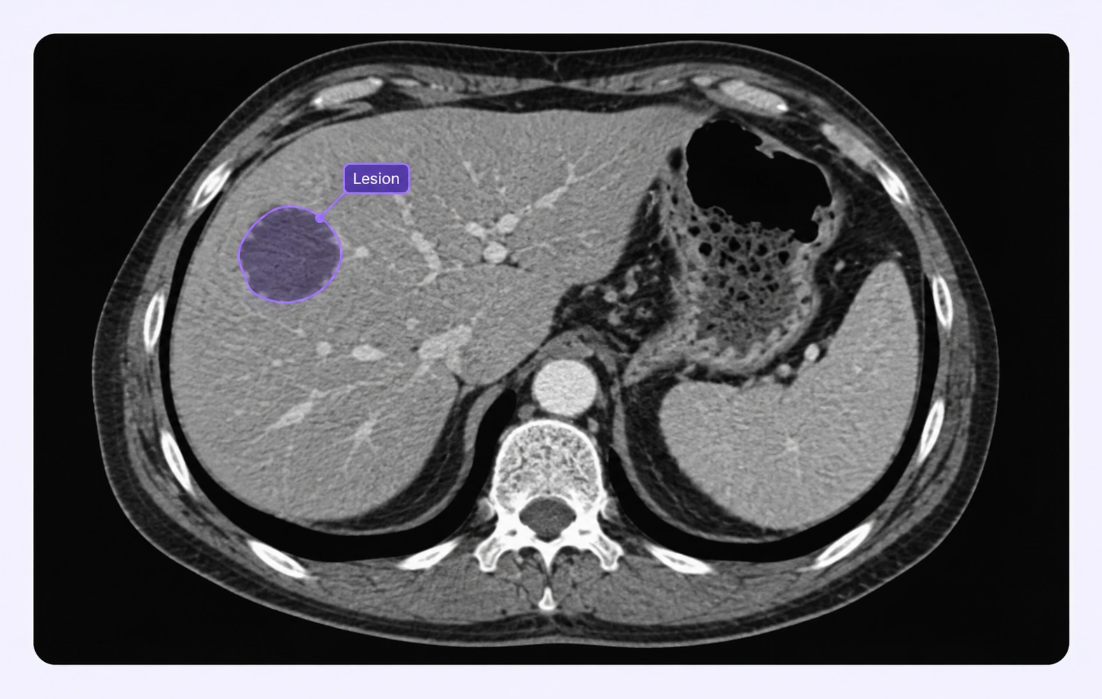
Trace the visible boundary of a lesion with a labeled contour under your task’s annotation criteria.
Create segmentation regions that describe the lesion class and retain the geometry needed by your training task.
It marks the regions belonging to a tumor or lesion so a supervised model can learn the boundaries specified by an expert annotation protocol.
Yes. Define the classes and properties required by your dataset, with clear rules for how annotators distinguish the categories.
Choose the geometry required by your model and annotation protocol. Contours express boundaries, while masks express the segmented region.
Yes. The medical viewer keeps labels in source image context, and supported volumes provide the available anatomical views for inspection.
The workflow creates and reviews lesion annotations for your downstream analysis. Measurement and response criteria should be defined and validated by your research team.
Document the boundary rule, flag uncertain regions with comments or properties, and use expert review to settle the final annotation.
Unitlab supports DICOM, NIfTI, and NRRD inputs. DICOM slices are grouped by series into an annotation volume, with available views determined by the imaging data.
Use a review stage to inspect labels against the source images, record comments, and return corrections through the workflow. Your team defines the annotation protocol and expert review criteria.
Yes. Versioned releases and supported exports let teams deliver reviewed annotations to downstream pipelines. Select an export that preserves the geometry and metadata your research task requires.


We use cookies to improve your experience. See our Privacy Policy and Cookie Policy.