
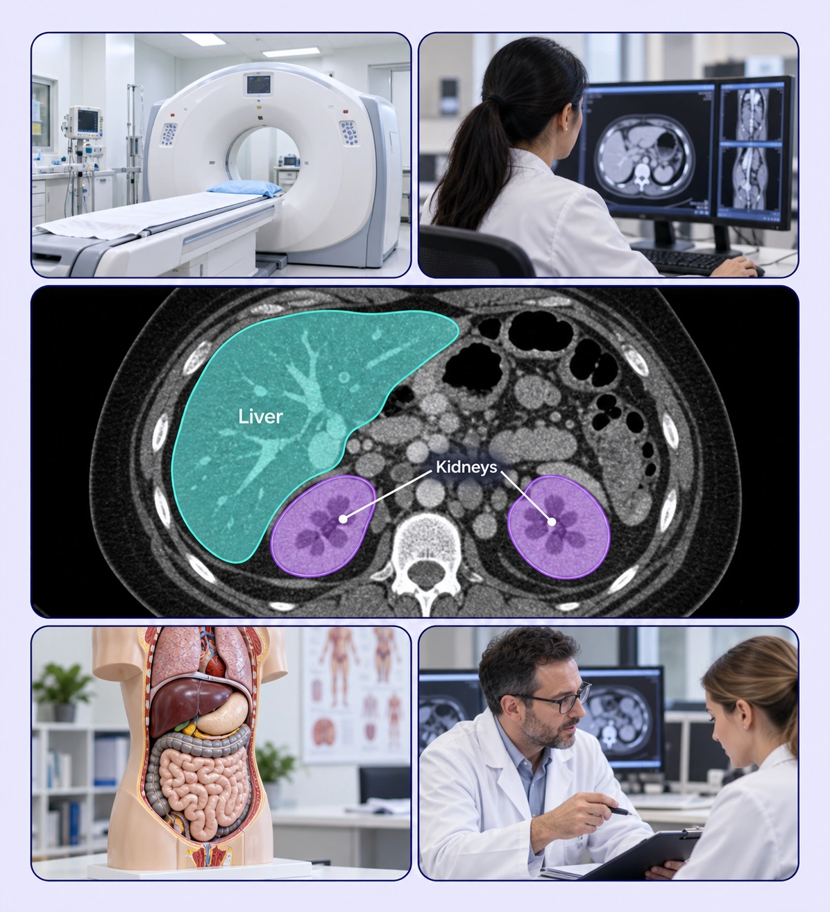
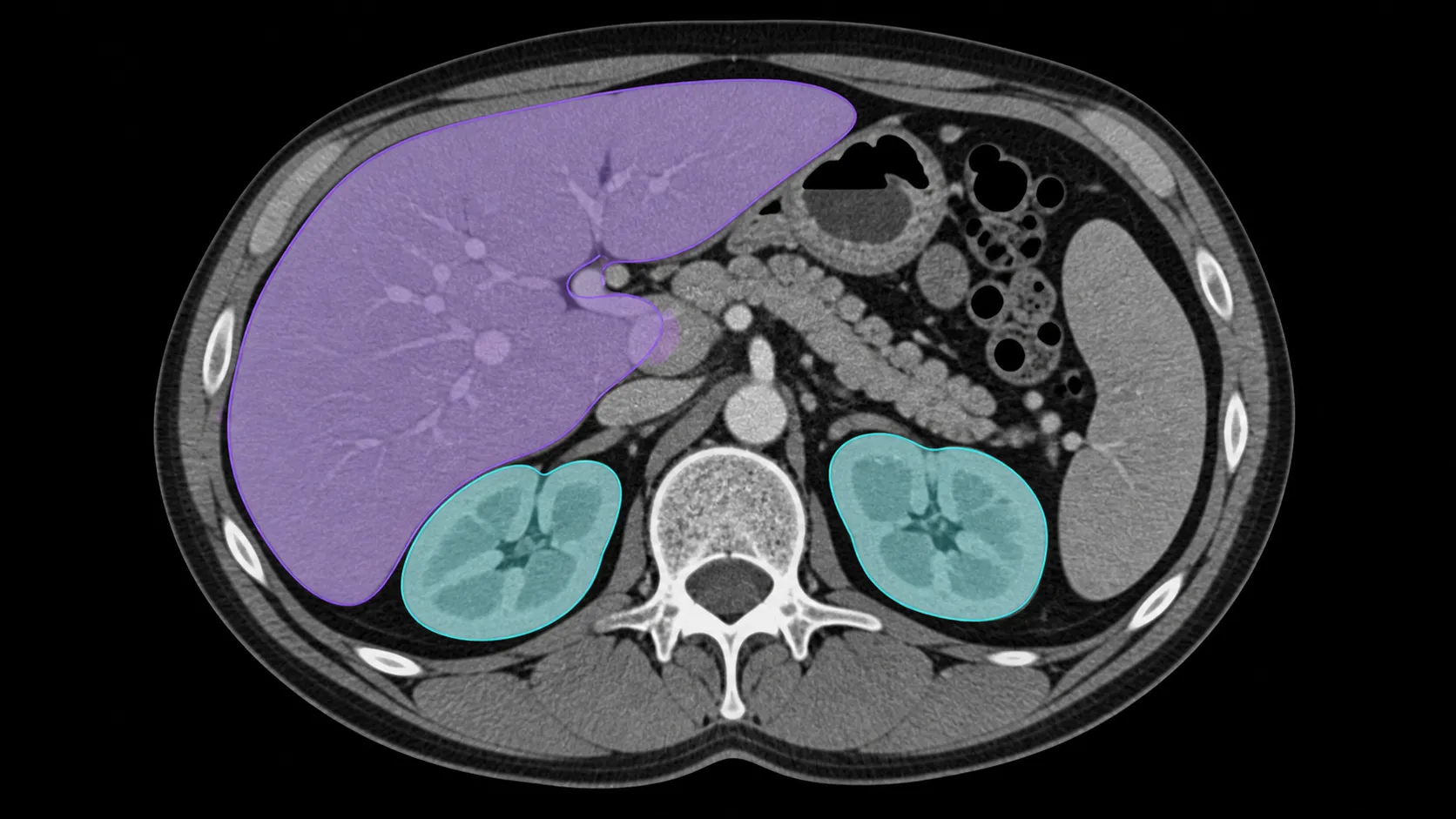
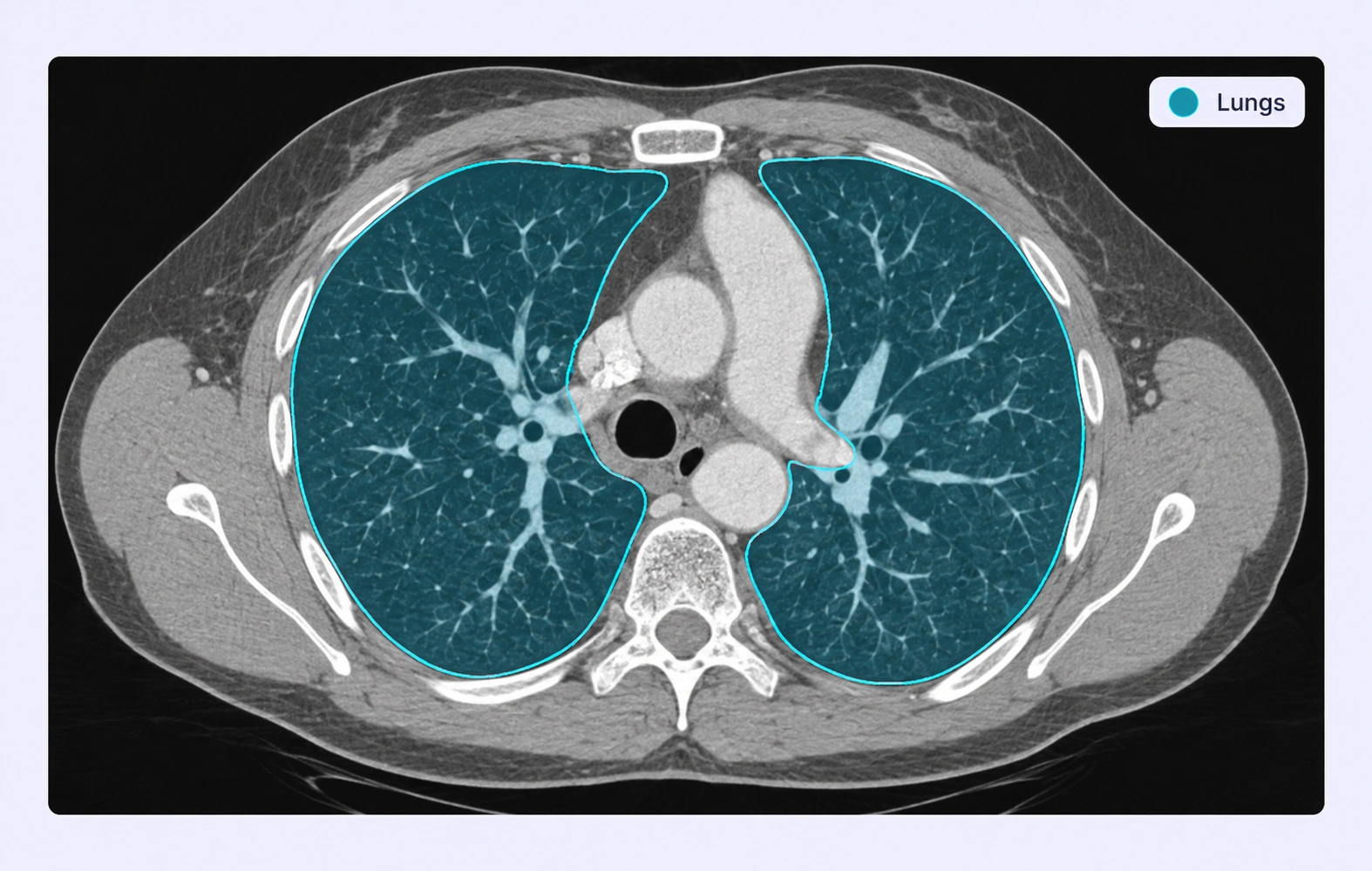
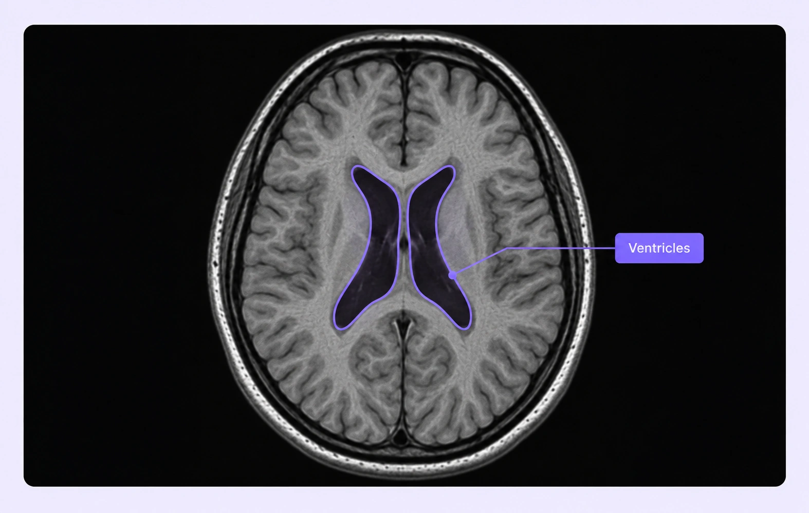
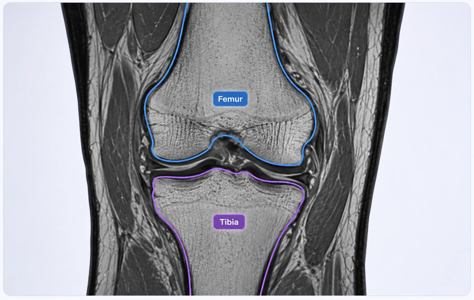
Segment organs and anatomy with model-assisted tools and expert review.
Automate curation, management, and versioning of anatomy datasets.
Reusable ontologies and governed quality control reduce labeling and review rework.
Mark the image regions that belong to each organ or structure using your project’s class definitions.
Use labeled contours for anatomy boundaries that your training pipeline represents as polygons.
Organ segmentation annotation marks anatomical regions in medical images, creating examples that teach a model to distinguish one structure from another.
Yes. Use separate classes for the structures required by your task and review how their boundaries relate in the source image.
Yes. Define anatomical classes and task-specific properties so the labeling schema follows your protocol.
Supported medical volumes expose available axial, sagittal, coronal, and 3D views. Use the views relevant to the structure and source data.
The tools can be used to mark visible structures at the scale of the source imaging data. Your protocol should define the resolution and boundary requirements.
Provide reference examples, define inclusion and exclusion rules, and have reviewers compare uncertain regions against the surrounding anatomy.
Unitlab supports DICOM, NIfTI, and NRRD inputs. DICOM slices are grouped by series into an annotation volume, with available views determined by the imaging data.
Use a review stage to inspect labels against the source images, record comments, and return corrections through the workflow. Your team defines the annotation protocol and expert review criteria.
Yes. Versioned releases and supported exports let teams deliver reviewed annotations to downstream pipelines. Select an export that preserves the geometry and metadata your research task requires.


We use cookies to improve your experience. See our Privacy Policy and Cookie Policy.